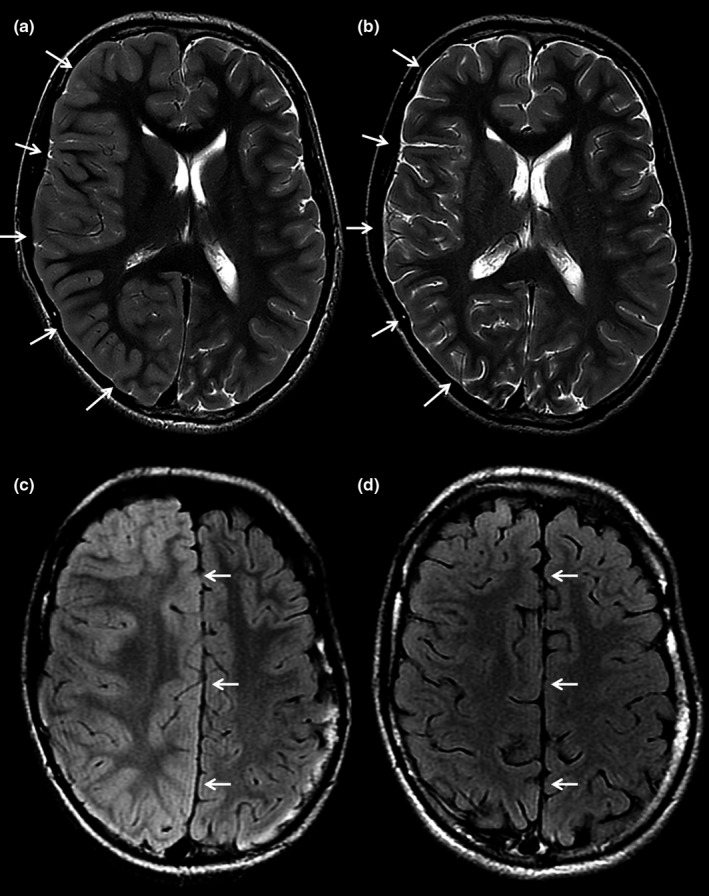
FIGURE 2.

Axial T2‐weighted (a) and T2 FLAIR (b) MR images of a patient a few hours after motor seizures affecting the left side of the body and follow‐up MR images four months later (c, d). The axial images at the level of the lateral ventricles (a) and centrum semiovale (b) show diffuse swelling of the cortex of the right cerebral hemisphere with obliteration of the sulci (arrows in a) and a midline shift due to the mass effect (arrows in b). The follow‐up images at the same brain levels demonstrate normalization of the cortical swelling and midline shift (arrows in c and d)
