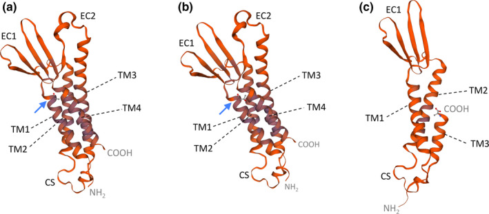
FIGURE 3.

Predicted 3D structures of wild‐type and mutant claudin‐16 proteins generated using homology modeling with SWISS‐MODEL.TM1 to 4, transmembrane domains 1 to 4; EC1 and EC2, extracellular loops 1 and 2; CS, cytoplasmic segment. (a) Wild type claudin‐16. (b) Mutant p.(Ala93Thr). Blue arrows indicate the location of the original alanine 93 residue and missense mutation p.(Ala93Thr). The NH2 and COOH termini correspond to threonine 70 and cysteine 255, respectively. (c) Deletion mutant lacking part of TM3, EC2, TM4, and COOH terminus. The COOH terminus corresponds now to alanine 197. The models are based on template 5b2 g.2 of claudin‐14
