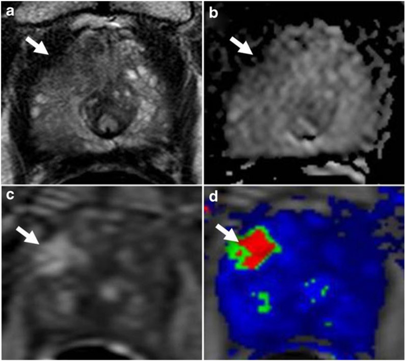
Fig. 1.
A 70 year old man with serum PSA of 51 ng/mL and four prior negative biopsies. (a) Axial T2W MRI, (b) ADC map from DW MRI, (c) raw DCE MRI , and (d) Ktrans map of DCE MRI depict a right apical-mid anterior transitional zone lesion (arrow). TRUS/MRI fusion-guided biopsy of the lesion revealed Gleason 4+ 4 tumor (100 % core involvement)

