Fig. 2.
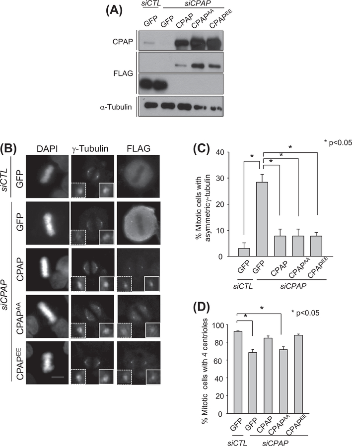
Rescue experiments with ectopic FLAG-CPAP proteins. (A) The CPAP-depleted HeLa cells were transfected with the FLAG-tagged expression vectors (pFLAG-GFP, pFLAG-CPAP, pFLAG-CPAPAA and pFLAG-CPAPEE). Expression of the endogenous and ectopic CPAP proteins was confirmed with the immunoblot analysis. (B) The CPAP-rescued cells were enriched at mitotic phase with a thymidine block and release, followed by the MG132 treatment for 1 h. The cells were immunostained with antibodies specific for FLAG and γ-tubulin. The scale bar represents 10 μm. (C) The mitotic cells with asymmetric distribution of γ-tubulin were determined by comparing relative γ-tubulin intensities between the spindle pole pair. More than 300 cells per group were analyzed from four independent experiments, and the results are presented as means and standard errors *P < 0.05. (D) The CPAP-rescued cells were immunostained with the antibodies specific to FLAG and centrin-2 to determine the centriole number at the mitotic spindle poles. For statistical analysis, more than 200 mitotic cells per group were analyzed from three independent experiments.

