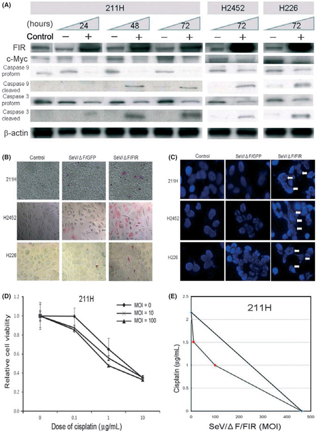
Fig. 2.
(A) Expression of FUSE-binding protein-interacting repressor (FIR), c-Myc, caspase 9 and caspase 3 after SeV/ΔF transduction. Lysate samples (20 μg/lane) from HMPM cells (211H, H2452 and H226) after fusion gene deleted non-transmissible Sendai virus vector encoding FIR (SeV/ΔF/FIR) transduction (+) at 100 MOI for 24, 48 and 72 h, medium only (control) and fusion gene deleted non-transmissible Sendai virus vector encoding green fluorescent protein (SeV/ΔF/GFP) (−) were subjected to western blot analysis. β-actin was used as the loading control. (B) APOPercentage Assay was used to detect early apoptosis in 211H, H2452 and H226 cells. More apoptotic cells were observed within 72 h after infection with SeV/ΔF/FIR at 100 multiplicity of infection (MOI) compared with SeV/ΔF/GFP and controls (PBS; original magnification, ×400). (C) DNA fragmentation as shown by DAPI stain. 211H, H2452 and H226 cells were infected with SeV/ΔF/FIR at 100 MOI, SeV/ΔF/GFP and controls (PBS). DAPI staining was performed 72 h after treatment with SeV/ΔF/FIR or SeV/ΔF/GFP. DNA fragmentation (arrows) was observed in SeV/ΔF/FIR-infected cells. (D) Synergistic activity of SeV/ΔF/FIR plus cisplatin in 211H cells, as shown by the leftward shift of the dose response curve of the MTS assay (error bars, SD). (E) Isobologram analysis (for ED50). The combined effects of SeV/ΔF/FIR and cisplatin were examined in 211H cells using at least two different dosing ratios.

