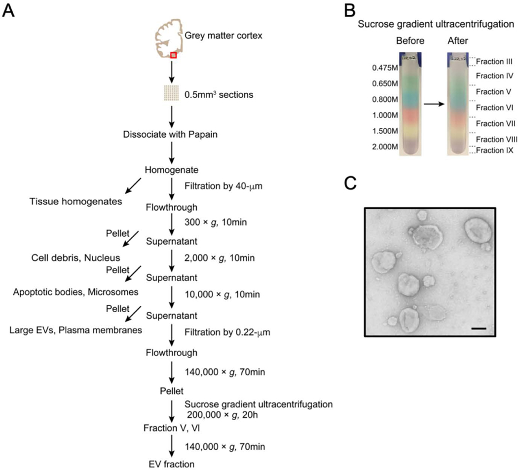
Figure 1. Flowchart of EV separation from unfixed human brain tissue via SG-UC method.
A) Sucrose gradient ultracentrifugation (SG-UC) protocol for separation of EVs from unfixed human frozen brain tissue. See section 3.1 for detailed method. Red square shows gray matter in brain cortex. B) Representative example of sucrose gradient before and after ultracentrifugation. C) Transmission electron microscopy (TEM) image of frozen human brain tissue-derived EVs (pooled fractions V and VI). Scale bar; 100 nm.

