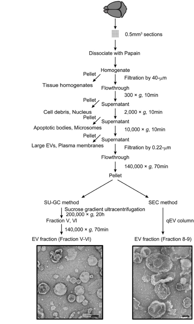
Figure 3. Flowchart of EV separation from unfixed murine brain tissue via SG-UC and SEC methods.
Sucrose gradient ultracentrifugation (SG-UC) or Size exclusion chromatography (SEC) protocols for separating of EVs from unfixed mouse frozen brain tissue. See section 3.1 for detailed methods. TEM image of frozen mouse brain-derived EVs (pooled fractions V and VI for SG-UC and pooled fraction 8 and 9 for SEC). Scale bar; 100 nm.

