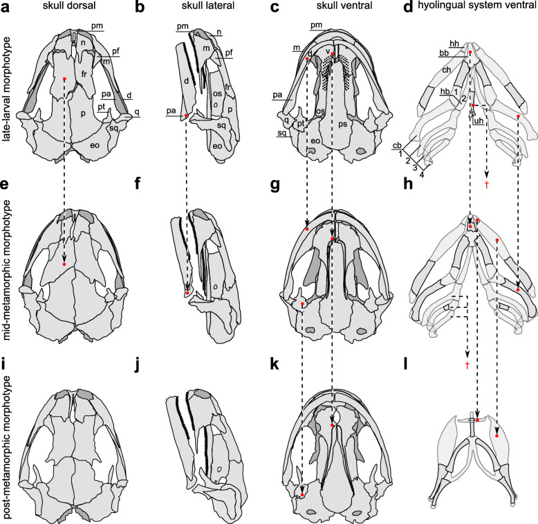
Fig. 1.
Skeletal morphology of the feeding apparatus of different morphotypes in I. alpestris. a-d (row 1) late-larval morphotype (LLM), e-h (row 2) mid-metamorphic morphotype (MMM), i-l (row 3) post-metamorphic morphotype (PMM). Abbreviations: (bb) basibranchial, (cb 1–4) ceratobranchial 1–4, (ch) ceratohyal, (d) dentary, (eo) exoccipital, (fr) frontal, (hh) hypohyal (also referred to as radial), (hb 1–2) hypobranchial 1–2, (m) maxilla, (n) nasal, (os) orbitosphenoid, (p) parietal, (pa) prearticular, (pf) prefrontal, (pm) premaxilla, (ps) parasphenoid, (pt) pterygoid, (q) quadrate, (sq) squamosal, (uh) urohyal, (v) vomer. Arrows connecting different morphotypes (rows) highlight significant structural differences. Arrows ending in the space between morphotypes marked with † indicate the reduction of the structure

