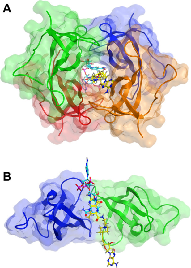
Figure 3.

Docking of 1 (yellow) into the DfrB1 tunnel with NADPH (cyan) (PDB: 2RK1). A pose with the adenosine moiety of 1 overlaying best with that of NADPH is shown (Figure S3). In the top 25 poses, inhibitor 1 forms contacts most frequently with K32, V66, and I68 (Figure 1; Figure S4). Contacts are rarely or not established with G35 and A36 that participate in binding the 2′-phosphate of NADPH, and with the YTT cluster.30 (A) Front view. (B) Side view; two subunits are represented.
