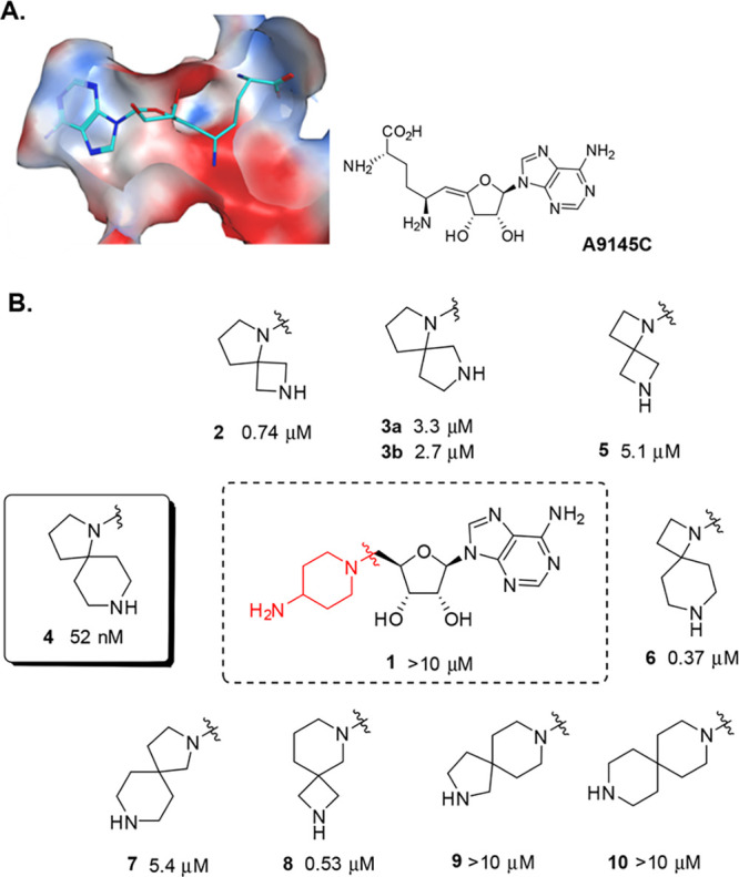
Figure 1.

(A) Molecular surface representation of PRMT5 bound to SAM analogue A9145C (cyan sticks; PDB 4GQB). The surface (clipped for clarity, front view is semitransparent) is colored by Poisson–Boltzmann electrostatics map ranging from −40 (red) through 0 (white) to +40 (blue) as modeled in Chemical Computing Group’s MOE software version 2016.0801. The histone H4 peptide is hidden to show pocket features near the SAM binding site. (B) Initial PRMT5 hits. Activities are IC50values in the enzyme assay. The absolute stereochemistry at the spiro diamine group in 3a/b is undefined, both diastereomers were prepared separately. For details, see SI, Section S2.
