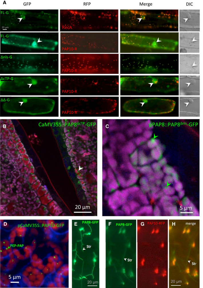
Figure EV2. (Related to Fig 3) Sub‐cellular localization of PAP8 variants.

-
ATransient assay in onion epidermal cells, pictures are given at the same scale. FL, full‐length ORF; −G, translational fusion with GFP; ΔNLS, deletion of the NLS; ΔcTP, deletion of the cTP, ΔΔ, deletion of both the NLS and the cTP. Onion cell co‐transfected with the corresponding variant fused to GFP and a plastid control fused to RFP (PAP10, PAP10‐RFP or RecA, RecA‐RFP) Merge, merged channels; DIC, differential interference contrast microscopy pictures to reveal the position of the nucleus within the cell when fluorescent nuclei were observed (marked with white arrowheads).
-
B, CConfocal imaging of stably expressed CaMV35S::PAP8∆cTP‐GFP (B) or pPAP8::PAP8∆nls‐GFP (C) in cotyledons of Arabidopsis thaliana; white arrowheads indicate nuclei; green arrowheads indicate sub‐plastidial localization. Observations similar to (C) were recorded for pPAP8::PAP8FL‐GFP.
-
DConfocal imaging of stably expressed CaMV35S::PAP10‐GFP in cotyledons of Arabidopsis thaliana showing the PEP‐PAP complex; the green arrowhead indicates the putative location of PEP‐PAP complexes within one chloroplast.
-
E–HPAP8‐GFP in stromules of onion epidermal cells expressed alone (E) or in co‐localization with PAP10‐GFP (F–H); stromules (str, white arrowheads) are only marked by PAP8‐GFP.
Source data are available online for this figure.
