
Figure 3. Astrocyte activation after microglial ablation.
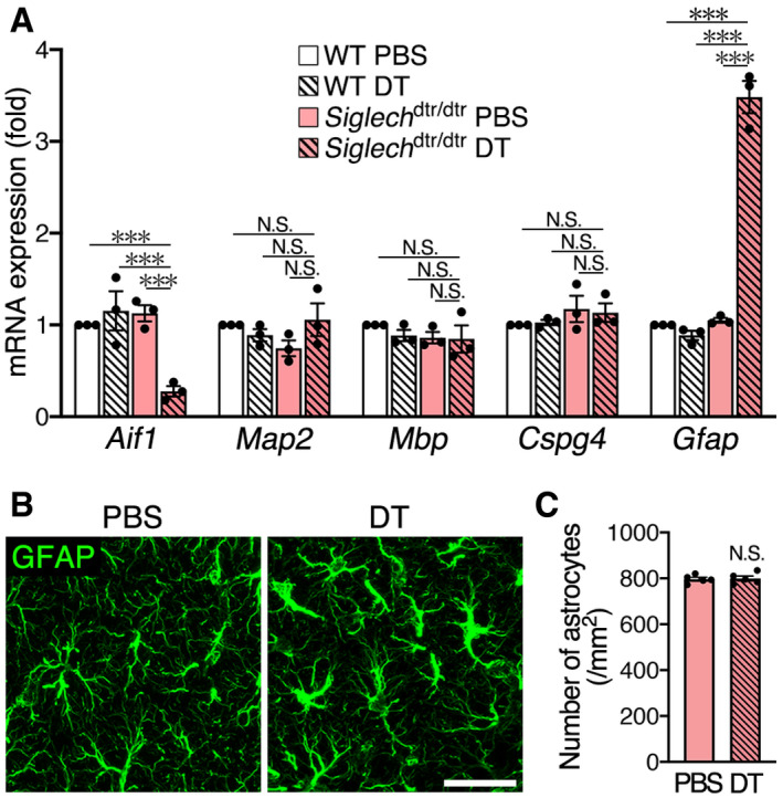
- qPCR analysis of marker molecules for CNS cell types. The hippocampus of WT and Siglech dtr/dtr mice 2 days after PBS or DT administration was analyzed (n = 3 animals per group, one‐way ANOVA with post hoc Tukey's test). Results are normalized to Gapdh and are shown as ratios to the value of WT mice injected with PBS.
- Immunohistochemical detection of astrocytes in Siglech dtr/dtr mice. Hippocampal CA1 sections were prepared 2 days after PBS or DT administration, and stained with an anti‐GFAP antibody. Images were acquired using the same laser power and sensitivity, and image processing was the same. Scale bar, 30 μm.
- Astrocyte number in hippocampal CA1 of Siglech dtr/dtr mice 2 days after PBS or DT administration (n = 5 animals, two‐tailed unpaired Student's t‐test).