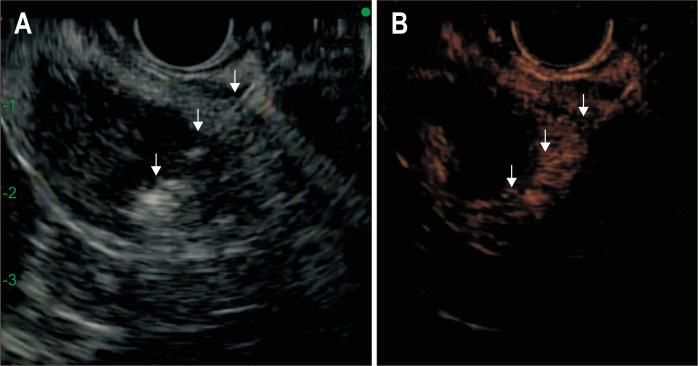
Fig. 3.
Conventional gray scale (A) and contrast-specific (ExPHD)-mode (B) during the second session of radiofrequency ablation. An arterial phase CEH-EUS image (B) obtained 5 days after RFA shows peripheral eccentric enhancement, suggesting a viable tumor. CEH-EUS facilitates the accurate targeting of the RFA needle (arrows) into the lesion to be treated.
CEH-EUS, contrast-enhanced harmonic endoscopic ultrasound; RFA, radiofrequency ablation.

