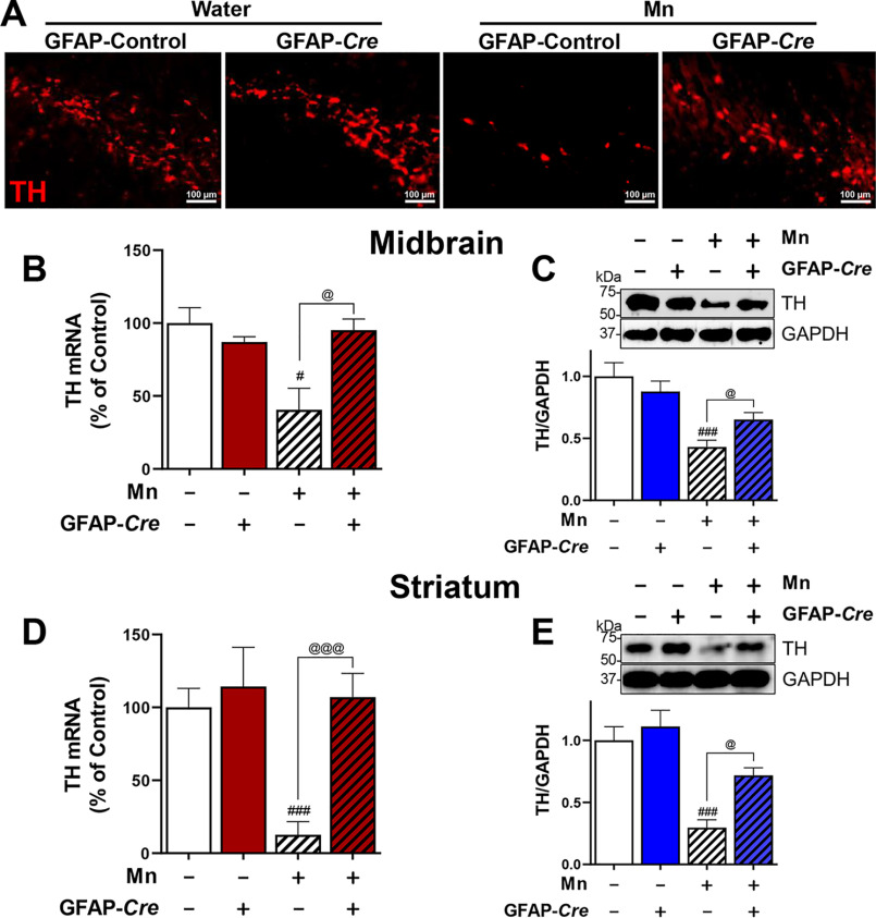
Figure 7.
Deletion of astrocytic YY1 attenuates Mn-induced decrease of TH expression in SN/midbrain and striatum. A, after Mn exposure (MnCl2, 30 mg/kg, intranasal instillation, daily for 3 weeks), mice were perfused, and coronal sections of brain tissues were immunostained with TH antibody as described under “Experimental procedures.” The expression of TH protein was shown as red fluorescence signals (TRITC) in the SN of the mouse brain (×10 magnification). B–E, after Mn treatment, midbrain (B and C) and striatal (D and E) regions were processed for TH mRNA and protein levels by qPCR and Western blotting, respectively, as described under “Experimental procedures.” GAPDH was used as a loading control. *, p < 0.05; #, p < 0.05; ###, p < 0.001 compared with GFAP-GFP/vehicle; @, p < 0.05; @@@, p < 0.001 compared with each other (one-way ANOVA followed by Tukey's post hoc test; n = 3). Data are expressed as mean ± S.D.

