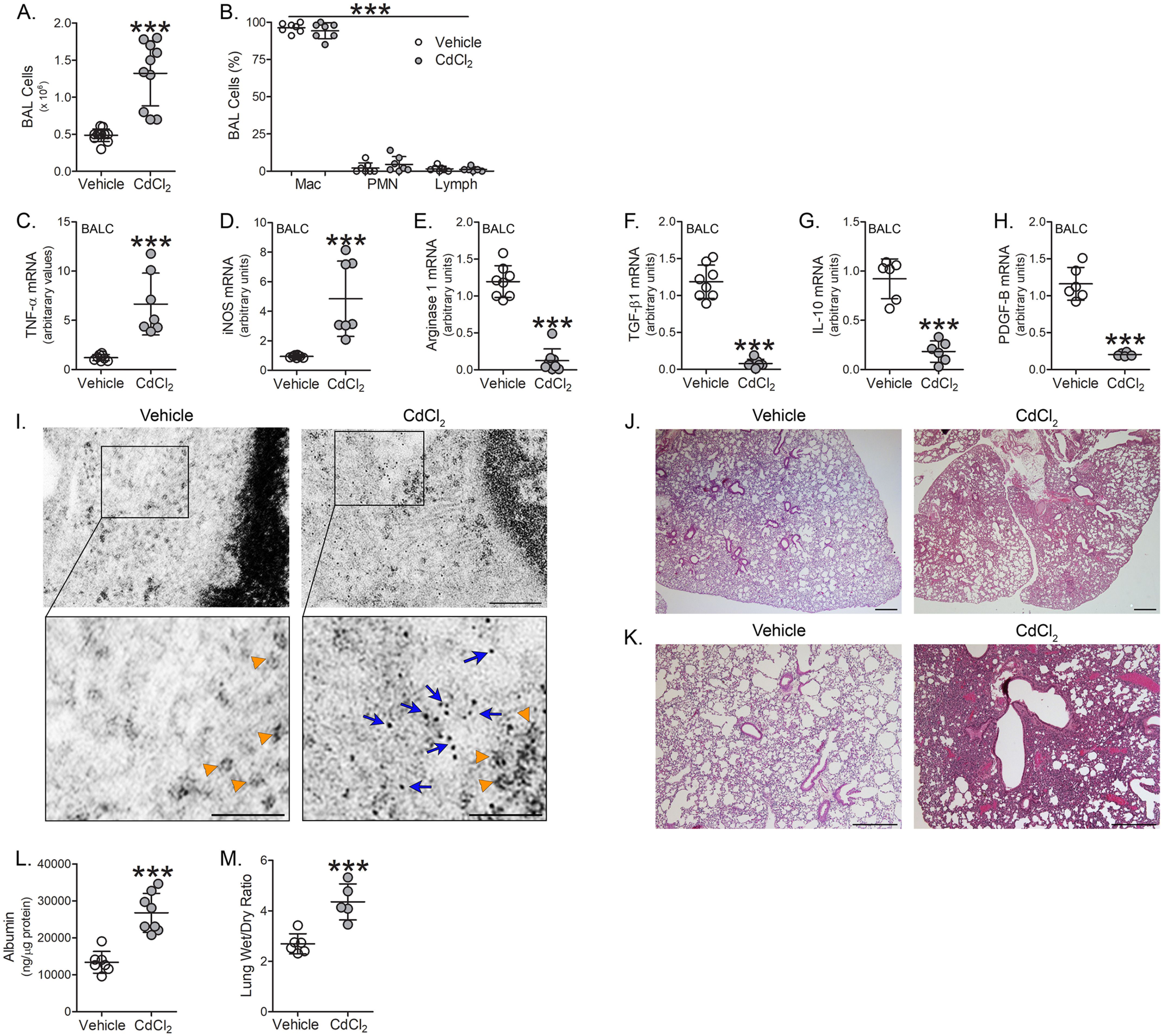
Figure 2.

Cadmium induces lung injury and the pro-inflammatory phenotype of lung macrophages. WT mice were exposed to vehicle or CdCl2 (100 ng/kg, intratracheal). After 7 days, BAL was performed. A, total number of BAL cells (n = 10-11) and B, cell differential (n = 7) from exposed mice. C, TNFα; D, iNOS; E, arginase 1; F, TGF-β1; G, IL-10; and H, PDGF-B mRNA expression in isolated BAL cells from exposed mice (n = 6-8). I, representative TEM analysis of BAL cells from exposed mice. Orange arrowheads indicate ribosomes. Blue arrows indicate cadmium particles. Scale bars: 200 nm (main), 100 nm (insets) (n = 3). J and K, representative H & E staining of lung tissue from exposed mice (n = 3). Scale bars: 500 nm. L, albumin levels in BAL fluid from exposed mice (n = 7-8). M, wet to dry ratio of lung weight from exposed mice (n = 5-6). p < 0.0001. Mac, macrophage; PMN, polymorphonuclear leukocyte; Lymph, lymphocyte. Values are shown as mean ± S.D.
