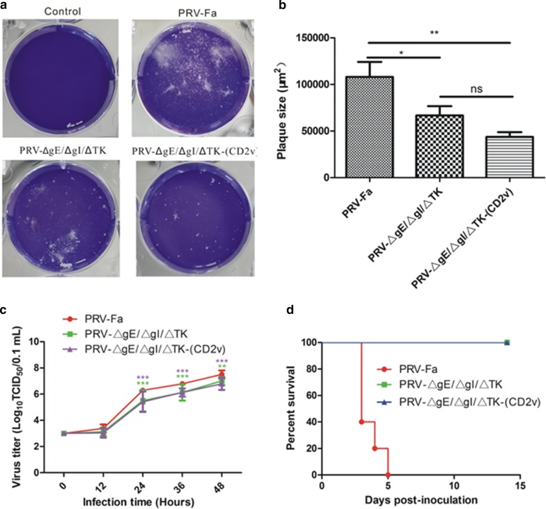
Fig. 2.
The insertion of CD2v did not change the proliferation and virulence of PRV-ΔgE/ΔgI/ΔTK. a The 50 TCID50 virus was inoculated into 5 × 10⁵ Vero cells and cultured for 48 h. The growth of plaques in Vero cells was observed by crystal violet staining. b Statistical results of the plaque area. c 1 × 10³ TCID50 viruses were inoculated into 5 × 10⁵ Vero cells to allow virus proliferation, and the virus sample was collected at 12 h, 24 h, 36 h, 48 h. The virus titer was calculated by the Karber method to draw a one-step growth curve. d Five-week-old SPF ICR mice were injected (i.m) with 5 × 10⁵ TCID50 viruses into the right hind leg to observe mice's survival daily, and the survival curve was drawn (n = 15/each group). Unpaired t-test or two-way ANOVA was performed by GraphPad Prism 5.0, GraphPad Software (San Diego, CA, USA), *p < 0.05, **p < 0.01, ***p < 0.001, ns (not significant)

