Fig. 2.
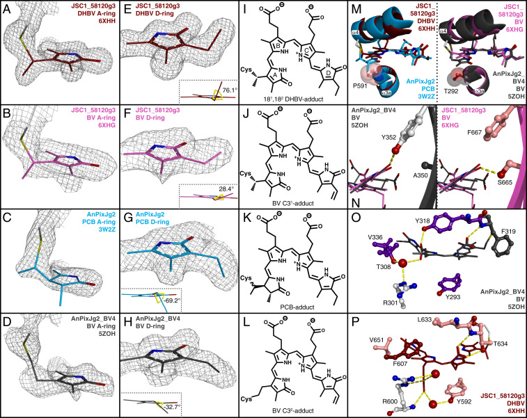
JSC1_58120g3 crystal structures. (A–D) Chromophore A-ring models and electron densities for JSC1_58120g3 coexpressed with PcyA (A) or with HY2 (B), PCB adduct of AnPixJg2 (C), and BV adduct of AnPixJg2_BV4 (D). (E–H) Chromophore D-ring models and electron densities are shown as in A–D. (Insets in E–H) C19-C18-C181-C182 dihedral angles. (I–L) Chemical representations of chromophore adducts as determined from A–H. (M) Alignments depicting helix α4 and chromophore position shift. Protein mainchain is depicted in ribbon view with Pro591 and Thr292 sidechains shown as space-filling spheres. JSC1_58120g3-DHBV is brick red, AnPixJg2 teal, JSC1_58120g3-BV magenta, and AnPixJg2_BV4 charcoal. DPYLoar-conserved residues are colored by atom with carbon in salmon. XRG-conserved residues are colored by atom with carbon in light gray. (N) Orientation of BV chromophore D-ring with XRG-conserved β6 residues (Left) or DPYLoar-conserved β6 residues (Right). (O) Chromophore arrangement and protein contacts in AnPixJg2_BV4. Colored as in M, and AmBV4-conserved residues are colored by atom with carbon in violet. Water molecules depicted as red spheres. (P) Chromophore arrangement and equivalent protein contacts in JSC1_58120g3-DHBV.

