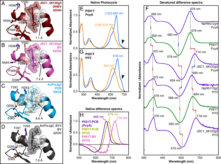
Fig. 3.
Phycobilin exclusion by Pro591. (A–D) View of the A-ring–binding pocket and nearby residues with opening width labeled for JSC1_58120g3-DHBV (A), JSC1_58120g3-BV (B), AnPixJg2 (C), and AnPixJg2_BV4 (D). Mainchain is depicted in ribbon view with Pro591/Thr292, chromophore, and primary cysteine represented as space-filling spheres. Coloring as in Fig. 2. (E) Dark-adapted 15Z (blue) and photoproduct 15E (orange) absorbance spectra are shown for P591T JSC1_58120g3 expressed with PcyA. Arrowhead denotes FR shoulder. (F) Stacked 15Z-15E difference spectra for denatured proteins compare P591T and WT JSC1_58120g3 to standards for PCB adduct (NpR6012g4) and PΦB adduct (NpR5113g2). (G) Spectra are shown for P591T JSC1_58120g3 expressed with HY2 as in E. (H) Native 15Z-15E difference spectra for P591T-PCB (violet) and estimated P591T-PΦB (bronze) and P591T-BV (pink) populations.

