Fig. 2.
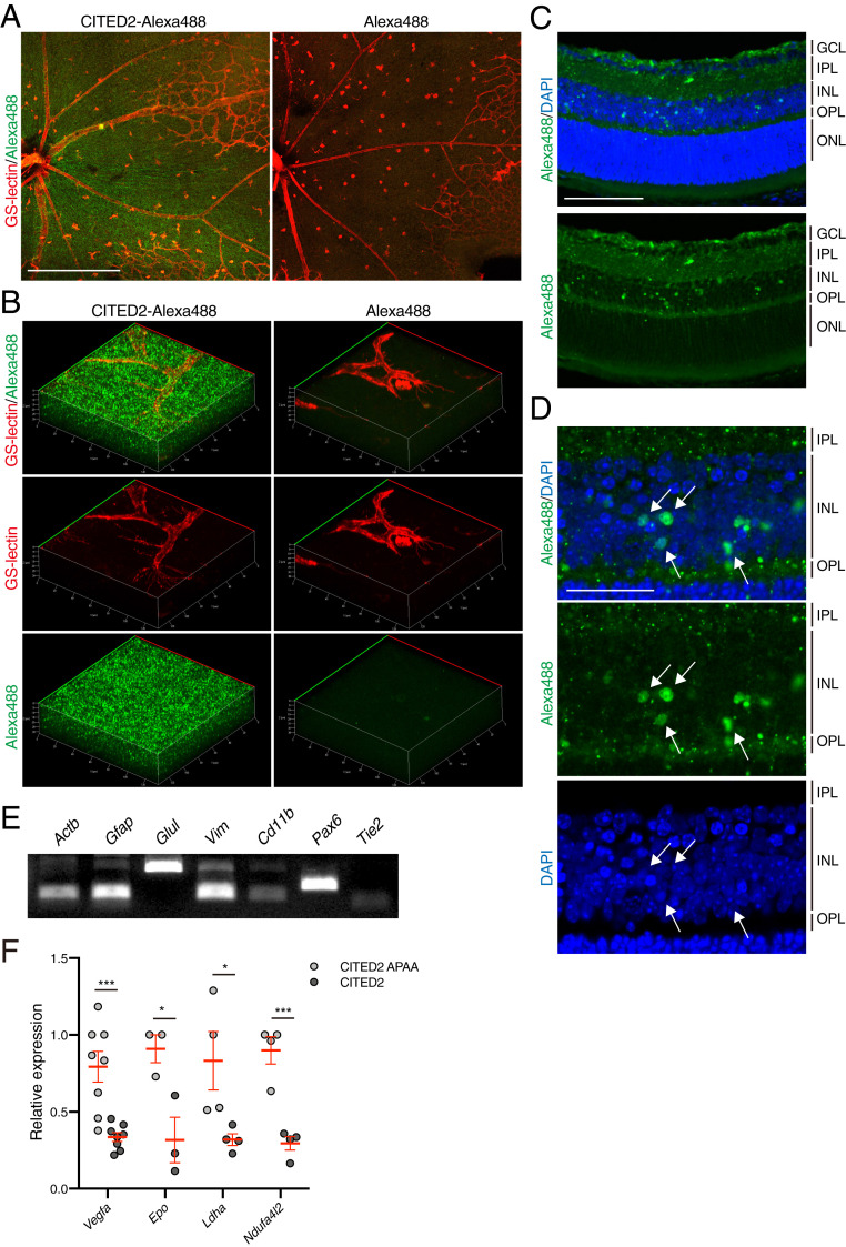
The CITED2 peptide is widely distributed in various retinal cell types after intravitreal injection. (A) Alexa488-CITED2 peptide fluorescence can be observed in OIR retinas. A total of 2 μM of Alexa488-conjugated CITED2 peptide (Left) and nonreactive Alexa488 dye (Right) were intravitreally injected into OIR mice on P12. Retinas were harvested 12 h after injection and retinal whole mounts were stained with GS-lectin (red). (Scale bars, 500 μm.) (B) Three-dimensional images of retinal whole mounts shown in A. (C) Representative cryosection of an Alexa488-CITED2 peptide-injected OIR retina. Retinal tissue was harvested 12 h after intravitreal injection on P12 and stained with DAPI. (Scale bars, 100 μm.) GCL, ganglion cell layer; IPL, inner plexiform layer; INL, inner nuclear layer; OPL, outer plexiform layer; ONL, outer nuclear layer. (D) High magnification images of the inner nuclear layer shown in C. White arrows highlight nuclear localization of CITED2 peptides. (Scale bars, 50 μm.) (E) Expression of retinal cell type-specific markers in Alexa488-CITED2 peptide-injected OIR retinas. OIR retinas were harvested and digested into a single cell suspension 12 h after intravitreal injection of Alexa488-CITED2 peptide on P12. Alexa488-positive cells were sorted and the expression of retinal cell markers was confirmed by RT-PCR. Actb, β-actin; Gfap, glial fibrillary acidic protein; Glul, glutamine synthetase; Vim, vimentin. (F) Expression levels of known HIF target genes in CITED2 peptide-containing retinal cells and CITED2 APAA peptide-containing retinal cells after intravitreal injections (n = 4 per sample). P values were calculated using multiple t tests. *P < 0.05, ***P < 0.001. The mean and SEM are shown in red.

