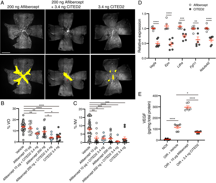
Fig. 5.
Combination therapy of the CITED2 peptide and aflibercept rescues retinal neovascularization and vaso-obliteration in OIR. (A) Immunofluorescent staining of OIR retinas at P17 after intravitreal injection of aflibercept (200 ng), aflibercept and CITED2 peptide (200 ng aflibercept and 3.4 ng CITED2 peptide), or CITED2 peptide (3.4 ng) at P12. Retinal whole mounts were stained with GS-lectin. Representative images for each treatment are shown (Upper) and the same images are shown (Lower) with NV highlighted in red and VO highlighted in yellow as used for quantification. (Scale bars, 1 mm.) (B) Quantification of the percentage of VO area in whole retinas. (C) Quantification of the percentage of NV area in whole retinas. For B and C, n > 7 per group. P values were calculated using one-way ANOVA with Tukey’s multiple comparisons test. *P < 0.05, **P < 0.01, ***P < 0.001, ****P < 0.0001. (D) Validation of expression of HIF target genes in CITED2- and aflibercept-treated OIR retinas. Total RNA was isolated 24 h after intravitreal injection of CITED2 peptide (3.4 ng) or aflibercept (10 μg) on P12 (n = 6 per group). P values were calculated using multiple t tests. **P < 0.01, ***P < 0.001, ****P < 0.0001. (E) Quantification of VEGF protein levels in P15 retinas by ELISA (n = 6 per group). P values were calculated using one-way ANOVA with Tukey’s multiple comparisons test. *P < 0.05, ****P < 0.0001. For B–E, the mean and SEM are shown in red.

