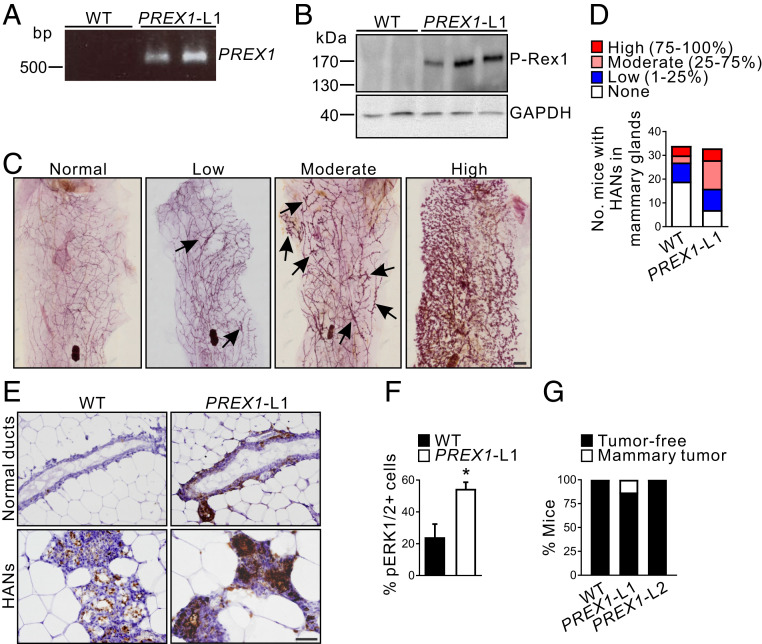
Fig. 1.
High P-Rex1 expression enhances hyperplastic alveolar nodule formation and induces de novo mammary tumor formation in aged mice. (A) Identification of WT and MMTV-PREX1 line 1 (PREX1-L1) transgenic mice by PCR of genomic DNA. (B) Whole-cell lysates from WT and MMTV-PREX1L1 transgenic mouse mammary epithelial cell organoids were immunoblotted with P-Rex1 or GAPDH antibodies. (C) Carmine alum-stained mammary gland whole mounts from 18- to 22-mo-old MMTV-PREX1L1 mice. Representative images of mammary glands with normal (0%), low (1 to 25%), moderate (25 to 75%), or high (75 to 100%) levels of HANs (arrows) are shown. (D) Data represent the number of 18- to 22-mo-old WT and MMTV-PREX1L1 mice with none, low, moderate, or high numbers of HANs in the mammary gland (WT n = 34, MMTV-PREX1L1 n = 33 mice). (E) Formalin-fixed, paraffin-embedded (FFPE) sections of mammary glands from 18- to 22-mo-old WT and MMTV-PREX1L1 mice were immunostained with pERK1/2 antibodies. (F) Quantitation of pERK1/2-positive epithelial cells in mammary ducts of 18- to 22-mo-old WT and MMTV-PREX1L1 mice. Data represent mean ± SEM (n = 5 mice/genotype, Student’s t test). (G) Tumor incidence in 18- to 22-mo-old WT and MMTV-PREX1 transgenic mice. Data represent the percentage of mice with or without mammary tumors (WT n = 50, MMTV-PREX1L1 n = 38, MMTV-PREX1L2 n = 19 mice). (Scale bars: 2 mm in C, 50 μm in E.) *P < 0.05.

