Fig. 4.
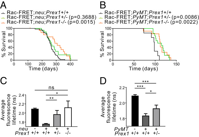
Prex1 ablation increases survival in oncogene-driven mouse models of breast cancer. (A and B) Kaplan–Meier survival curves of (A) Rac1-FRET;neu (n = 45), Rac1-FRET;neu;Prex1+/− (n = 18), Rac1-FRET;neu;Prex1−/− (n = 16) mice, and (B) Rac1-FRET;PyMT (n = 7), Rac1-FRET;PyMT;Prex1+/− (n = 13), Rac1-FRET;PyMT;Prex1−/− (n = 12) mice showing Prex1 loss significantly increases survival to endpoint (primary tumor ≥1.5 cm diameter) (Log-rank Mantel–Cox test). (C–D) Rac1-FRET;neu;Prex1 (C) or Rac1-FRET;PyMT;Prex1 (D) mice at clinical endpoint were imaged on a multiphoton system. Quantitation of average fluorescence lifetimes of individual cells inside the primary tumor mass at clinical endpoint. Data represent mean fluorescence lifetime ± SD [n = 3 mice/genotype, (C) 257 cells, (D) 264 cells] (one-way ANOVA and Tukey’s multiple comparisons test). *P < 0.05, **P < 0.01, ***P < 0.001, ns, not significant.

