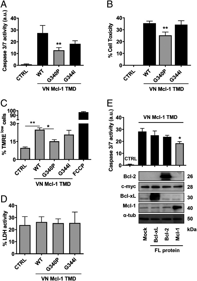
Fig. 2.
Mcl-1 TMD induces cell death. (A) Caspase 3/7 activity and cell toxicity (B) induced by the VN Mcl-1 WT TMD and single amino acid mutants were analyzed in cytosolic extracts of HCT116 cells 16 h after transfection. CTRL means nontransfected cells. Error bars represent the mean ± SEM, n = 3. P value, according to Dunnett’s test, displayed. **P < 0.01. Mitochondrial polarization status (C) and LDH activity (D) in the above-described conditions. The mitochondrial uncoupler FCCP was used as a positive control for mitochondrial depolarization. (E) HCT116 cells were transfected with Mcl-1, Bcl-xL, or Bcl-2 FL proteins. After 24 h, the VN Mcl-1 TMD was expressed for 16 h, and caspase 3/7 activity measured in cytosolic extracts. CTRL refers to nontransfected cells and mock condition refers to cells transfected with the corresponding empty vector for FL expression proteins. Error bars represent the mean ± SEM, n = 5. *P < 0.05. Chimeric protein expression of the VN Mcl-1 TMD (c-myc) and Mcl-1, Bcl-xL or Bcl-2 FL proteins are shown in the Bottom using α-tubulin as a loading control.

