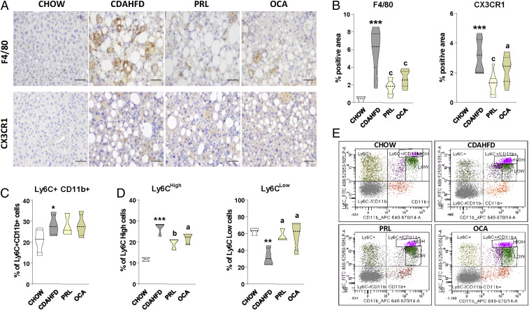
Fig. 3.
Effects of PRL on liver and peripheral blood monocytes. (A) Representative photomicrographs (magnification 200×) of liver sections stained with specific F4/80 and CX3CR1 antibodies used for the assessment of total liver macrophages and infiltrated liver macrophages, respectively. (Scale bars, 50 μm.) (B) Histomorphometric analysis of the area stained with F4/80 and CX3CR1 antibodies. (C) Percentage of total monocytes in the peripheral blood of mice determined by flow cytometry (n = 4 for each group). (D) Percentage of Ly6CHigh and Ly6CLow monocytes among the Ly6C+Cd11b+ cells. (E) Representative flow cytometry plots for Ly6C-expressing monocytes. Results are expressed as mean ± SEM. *P < 0.05; **P < 0.005 and ***P < 0.001 vs. chow; aP < 0.05, bP < 0.005, and cP < 0.001 vs. CDAHFD.

