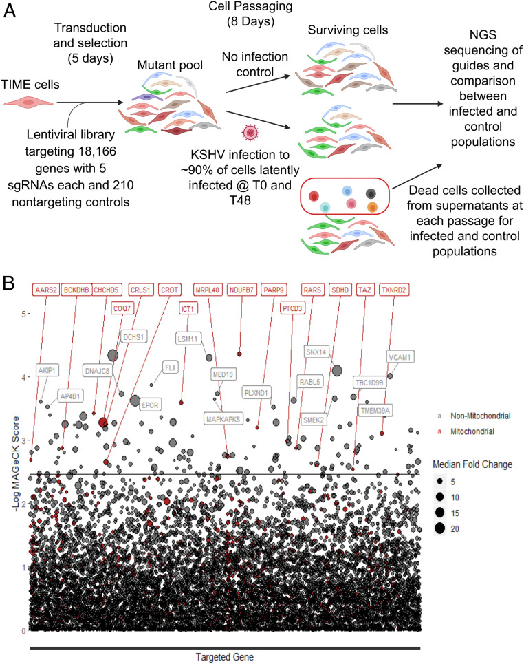
Fig. 1.
CRISPR-Cas9 whole-genome screen to identify essential host factors during KSHV infection of endothelial cells. (A) Schematic of TIME cell whole-genome screen of KSHV-infected cells (T0 and T48 refer to zero and 48 hours post initial KSHV infection, respectively. NGS stands for next generation sequencing). (B) Plot of the results of the live cell screen. The back line represents the false discovery rate cutoff of 0.25. The size of the circles represents the magnitude of the median log fold change for all sgRNAs for that particular gene. All red circles accompanied by red text are genes whose gene products localize to mitochondria.

