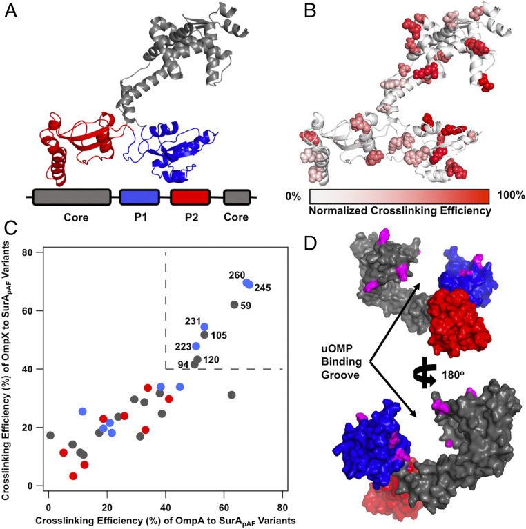
Fig. 1.
Open SurA binds client uOMPs in a groove between domains. (A) The structure of open SurA shown as a schematic, with the domains colored as depicted in the sequence diagram below (core, gray; P1, blue; P2, red). In this conformation, the three domains of SurA are structurally isolated from each other and do not form extensive interdomain contacts. (B) The 32 surface exposed sites on SurA in which pAF was substituted, shown in a space-filling representation. Photo-crosslinking was induced with 5-min UV exposure. Each crosslinking site is colored based on the normalized crosslinking efficiency to uOmpA171 as based on quantitative SDS/PAGE (SI Appendix, Fig. S2). The highest crosslinking sites are found on the core and P1 domains of SurA while P2 exhibits only modest crosslinking efficiency. (C) The raw crosslinking efficiencies of SurA to uOmpA171 and uOmpX are shown and colored by the SurA domain in which they residue (as in A). Eight SurApAF variants stand out by having high (>40%) crosslinking efficiency to both uOMP clients and are labeled with their residue number in the upper right quadrant of the graph (demarcated by dotted lines). (D) The eight high efficiency crosslinking sites, shown in magenta, are mapped onto a surface representation of the structure of open SurA. Together, these sites line a groove formed between the core and P1 domains, indicating that uOMPs are primarily bound there.

