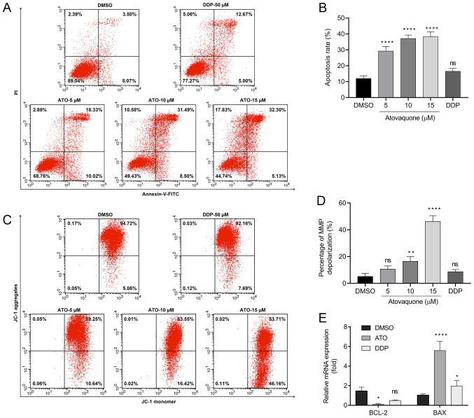
Figure 3.
ATO induces apoptosis of EpCAM+CD44+ HCT-116 cells in hypoxia. (A and B) Flow cytometric analysis of Annexin V/PI staining of EpCAM+CD44+ HCT-116 cells treated with ATO (5, 10 and 15 µM) under hypoxic conditions. (A) Flow cytometry dot plots of cells with Annexin V/FITC and PI fluorescence staining. Apoptotic cells are contained in the lower-right and upper-right quadrants. (B) Quantification of apoptotic cells. (C) Flow cytometric analysis of JC-1 staining assay of EpCAM+CD44+ HCT-116 cells treated with ATO (5, 10 and 15 µM) under hypoxic conditions. (D) Percentage of MMP depolarization. (E) Reverse transcription-quantitative PCR analysis of Bcl-2 and Bax mRNA expression in EpCAM+CD44+ HCT-116 cells treated with ATO (15 µM) under hypoxia. Cells treated with 50 µM DDP were used as the positive control. The relative mRNA expression is presented relative to the DMSO control. Values are expressed as the mean ± standard deviation of three independent experiments. *P<0.05, and ****P<0.0001 vs. DMSO as determined by one-way ANOVA by following Dunnett's test. ATO, atovaquone; DDP, cisplatin; ns, no significance; EpCAM, epithelial cell adhesion molecule; MMP, mitochondrial membrane potential.

