Fig. 5.
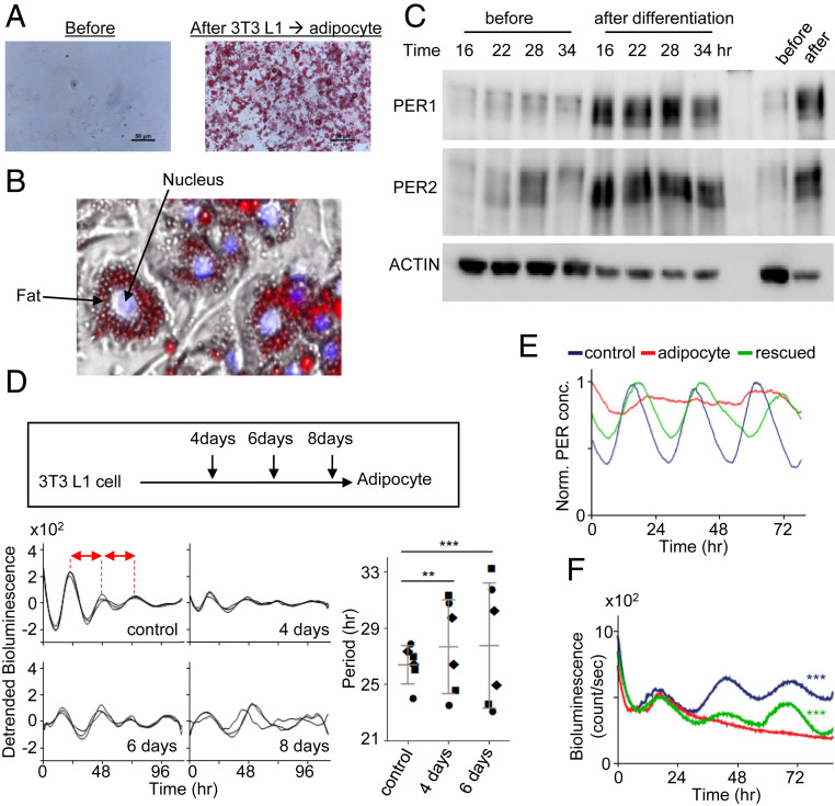
Spatial regulation of PER is directly linked to temporal manifestation of PER and circadian rhythms. (A and B) Fat vacuoles (Oil Red O) were enriched in the cytoplasm of adipocytes differentiated from 3T3 L1 cells. A zoomed-in image from the right image in A is shown in B. (C) Both hypophosphorylated and hyperphosphorylated PER species were visible at all times in the adipocytes. Unsynchronized cells are shown in the last two lanes. (D) Circadian rhythms were gradually compromised as L1 cells were differentiated into adipocytes and completely lost on day 8. Note that circadian period is unstable and generally longer in partially differentiated day 4 and day 6 cells compared with controls. The periods were estimated by peak-to-peak (red arrows) analysis in the samples. Three different symbols represent three independent samples. Data are mean ± SD, representative of two experiments. (E) Our model predicts that arrhythmicity in adipocytes can be rescued by increasing Per promoter activity. (F) Robust bioluminescence rhythms were recovered in adipocytes when PER2 was overexpressed using an adenoviral vector-delivered Per2 transgene (Per2 promoter and Per2 coding sequence). n = 3 each. Data are representative of two experiments.

