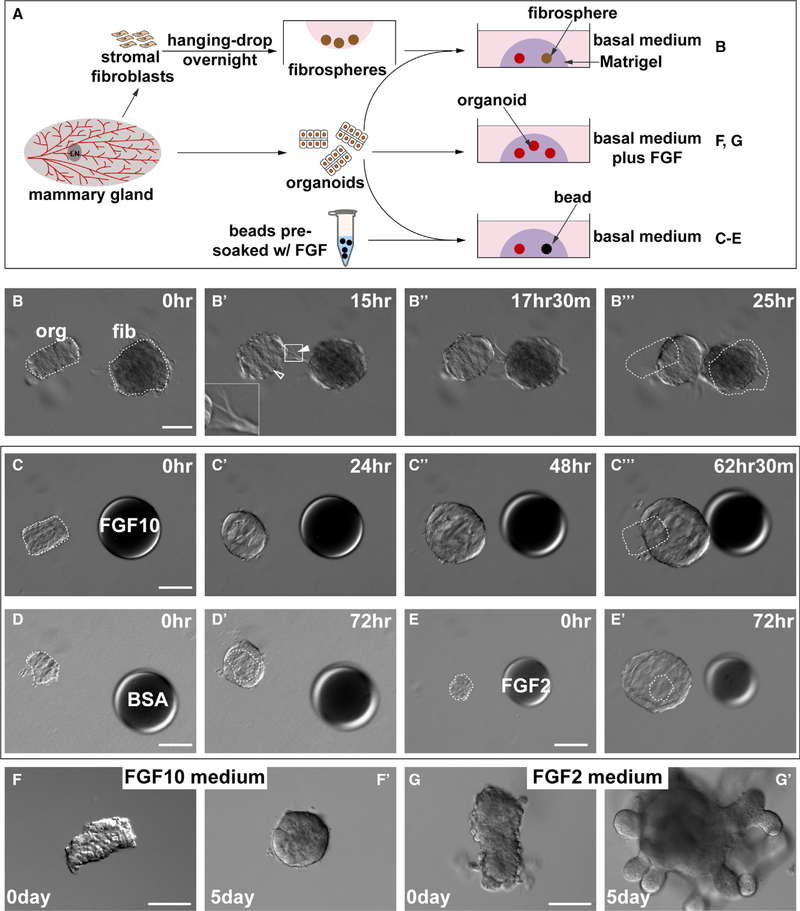
Figure 1. Mammary Gland Epithelium Undergoes Directional Migration toward the FGF10 Signal.
(A) A schematic diagram depicts the experimental procedures during sample preparation, in vitro culture, and treatment methods. Mammary organoids and stromal fibroblasts were prepared from wild-type mice. Heparan sulfate beads were pre-soaked in fibroblast growth factors (FGFs) overnight and were briefly rinsed before use. Organoids were co-cultured with aggregated fibroblasts, the “stromospheres” in (B)–(B″′) and the beads pre-soaked in FGFs or BSA in (C)–(E) or in medium containing FGFs in (F)–(G′).
(B) Time course of in vitro co-cultures of epithelial organoids with stromospheres. Note the epithelium formed a cyst (hollow arrowhead) and underwent directional collective migration toward the stromospheres, whereas the stromospheres formed spiky extensions made of fibroblasts (filled arrowheads), often toward the direction of the epithelium (n = 5). White dotted outlines indicate the original positions of the organoid and stromosphere at time 0 h. Scale bars, 100 μm.
(C–E) Differential responses of epithelial organoids to beads pre-soaked in FGF10 (C), BSA (D), or FGF2 (E). (C)–(C″′) Time course of directional migration of epithelial organoid toward the FGF10 bead (n = 23). Organoid did not migrate toward the beads pre-soaked in BSA (D) and (D′) (n = 37) or FGF2 (E) and (E′), n = 7). Instead, when stimulated by FGF2, organoids formed huge cysts. Heparan acrylic beads of ~100 μm in diameter were juxtaposed with mammary organoids at a distance of ~100 μm. Scale bars, 100 μm.
(F and G) Epithelial organoid response to FGF10 (F) and (F′) (n = 5) or FGF2 (G) and (G′) (n = 33) when it was universally delivered in the medium. Note organoid epithelium formed branches after 5 days of culture in a medium containing FGF2 (G) and (G′). Scale bars, 100 μm.

