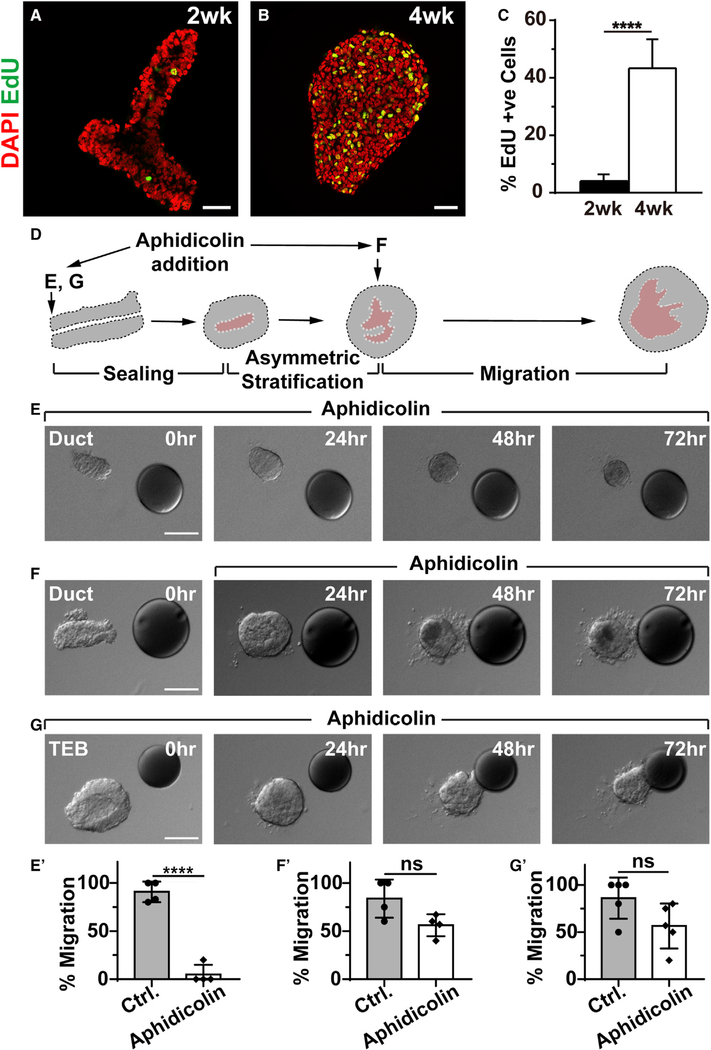
Figure 3. Frontal Epithelial Stratification Is Required for Collective Directional Migration.
(A–C) Cell proliferation as examined by EdU (red) incorporation during the transformation of the mammary epithelium from a simple ductal epithelial end at the prepubertal (2 weeks, n = 7) stage (A) into a bulb-shaped stratified end bud at the pubertal (4 weeks, n = 4) stage (B). Quantification of cell proliferation (C). Scale bars, 30 μm.
(D–G) Time course of epithelial organoid migration toward the FGF10 beads in the presence of cell proliferation inhibitor aphidicolin to block stratification. (D) Diagram illustrating the drug-treatment regimens. Aphidicolin was applied to the organoid either at the beginning, when it was a simple ductal epithelium (E) (n = 20), or after stratification was complete (F) (n = 20). Alternatively, the inhibitor was given to the organoid in the form of a TEB, which was a stratified epithelium (G) (n = 25). Scale bars, 50 μm. (E′)–(G′) Quantification of the percentages of organoids that migrated under the above treatment strategies. ****p < 0.0001. ns, not significant.

