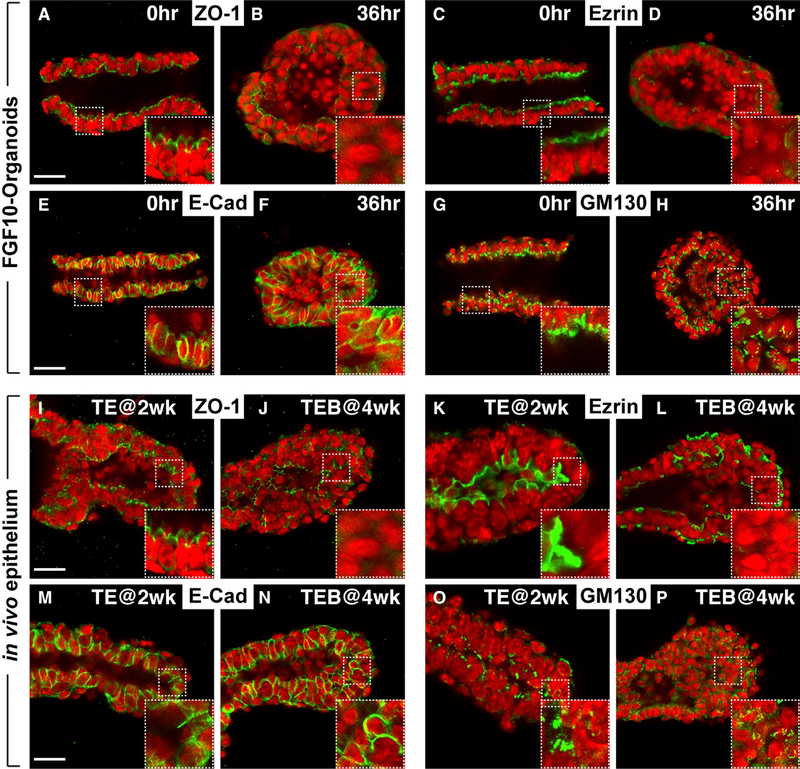
Figure 4. Apical-Basal Polarity Is Lost in Stratified Epithelium Both In Vitro and In Vivo.
(A–H) Immunofluorescence examination of tissue polarity markers in organoids at the duct (A, C, E, and G) and at the migrating cyst stages (B, D, F, and H). Insets are close-up views of the area in the white rectangles with dashed lines. Note ZO1 and Ezrin are normally expressed in the apical membrane (A) (n = 15) and (C) (n = 6) but are lost in cells of the stratified epithelium (B) (n = 5) and (D) (n = 9), except in those facing the lumen. E-cadherin is normally present in the basal-lateral membrane of simple ducts (E) (n = 9) but are found throughout the membrane of stratified epithelial cells (F) (n = 5). GM130 is often found toward the apical side in ductal epithelium (G) (n = 10), but that orientation becomes random in migrating organoids (H) (n = 5). Scale bars, 20 μm.
(I–P) Immunofluorescence examination of the above tissue polarity markers in vivo in the ductal epithelial TEs at prepubertal (2 weeks): (I) (n = 6); (K) (n = 3); (M) (n = 5); and (O) (n = 4) and in the TEBs at pubertal stages (4 weeks): (J) (n = 3); (L) (n = 3); (N) (n = 4); and (P) (n = 3). Insets are close-up views of the area in the white rectangles with dashed lines. Note the expression patterns of in vivo TEBs (J, L, N, and P) are similar to those of the migrating cysts in vitro (B, D, F, and H). Scale bars, 20 μm.

