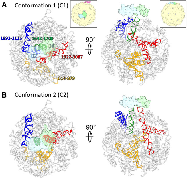
FIGURE 6.

Two conformations of the gRNA inside MS2. (A,B) Two dominant gRNA conformations of the MS2 (C1,C2) are modeled based on the two cryo-EM maps. The gRNA fragments with large differences between the two models are colored yellow (gRNA fragment 614–879), green (gRNA fragment 1643–1700), blue (gRNA fragment 1992–2125), and red (gRNA fragment 2922–3087). Two copies of the coat protein dimers, which switch interacting stem–loop partners, are outlined by transparent green (Dimer 1: D1) and transparent blue (Dimer 2: D2). The insets of the capsids (magenta: maturation protein, green: D1, blue: D2, yellow: rest of the coat protein shell) show the viewing orientation for the corresponding columns and the location of the two dimers on the capsid.
