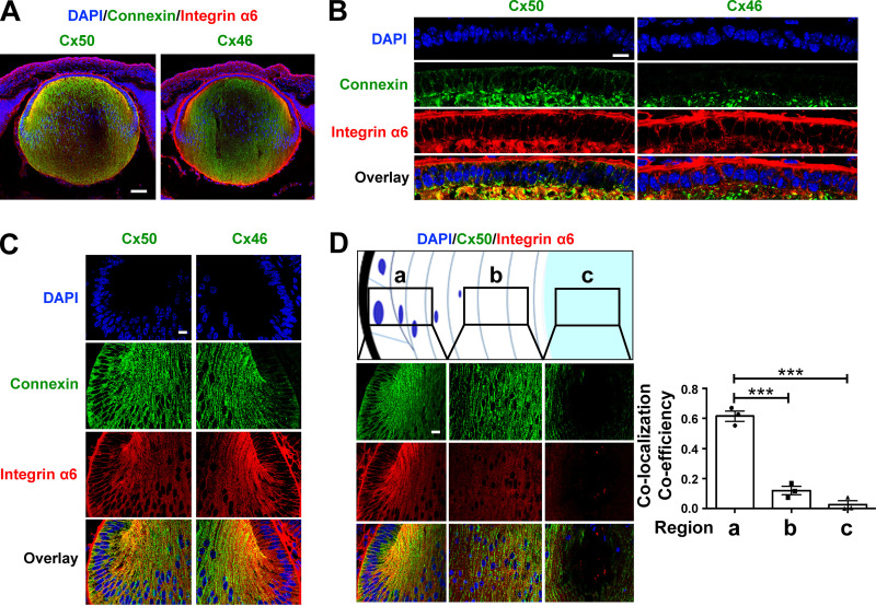
Figure 2.
Cx50 colocalized with integrin α6 in lens epithelium and outer cortical fiber cells. (A) Embryonic day 18 mouse lens sagittal cryosections were immunostained with anti-Cx46 (green), Cx50 (green), or integrin α6 (red) antibody and counterstained with DAPI (blue), followed by corresponding secondary antibodies conjugated with either Alexa Fluor 488 or rhodamine. The corresponding images were merged (Overlay). Scale bar, 100 µm. (B and C) Anterior epithelial cells (B) and equatorial epithelial and nascent fiber cells (C). Scale bar, 20 µm. (D) Different depths of the lens regions from equator as illustrated in panels a (equator), b (cortical), and c (nucleus), Cx50 and integrin α6 were double immunostained (left), and the extent of colocalization was quantified and presented as colocalization coefficiency using ImageJ (right). Thick black outline indicates the lens capsule. Scale bar, 20 µm. Each data point in the graph represents an individual mouse in each group. The data are presented as mean ± SEM. ***, P < 0.001.

