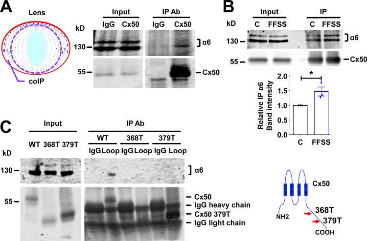
Figure 5.
Integrin α6 associates with lens Cx50. (A) Crude membrane extracts of cortical fiber portions of E19 chick lens as illustrated in the diagram (left) were collected and immunoprecipitated with control IgG or anti-Cx50 CT antibody. Preloading lysate (Input) and immunoprecipitates (IP) were immunoblotted with anti-integrin α6 and anti-Cx50 loop domain antibody (right). (B) CEF cells infected with RCAS(A) Cx50 were subjected to FFSS at 1 dyn/cm2 for 30 min (FFSS) or under static conditions (C) and immunoprecipitated with anti-FLAG antibody. The immunoprecipitates were immunoblotted with anti-integrin α6 and anti-Cx50 CT antibody (upper panel). Relative immunoprecipitated integrin α6 to immunoprecipitated Cx50 band intensity was quantified (lower panel). Each data point in the graph represents an individual experiment out of three repeats (n = 3). (C) CEF cells infected with RCAS(A) Cx50 and Cx50 mutants 368T and 379T. Crude cell membrane extracts were immunoprecipitated with IgG control or anti-Cx50 intracellular loop domain antibody. The immunoprecipitates were immunoblotted with anti-integrin α6 or anti-Cx50 loop domain antibody (left). The truncation sites at C-terminus of Cx50 are indicated (right). Data are presented as mean ± SEM. *, P < 0.05.

