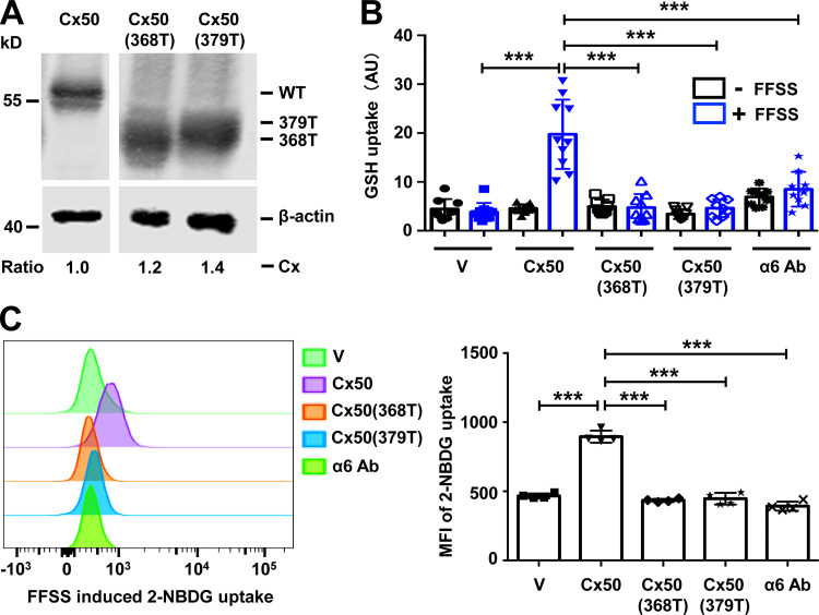
Figure 7.
Association of integrin α6 and Cx50 is essential for the permeability of glucose and GSH through hemichannels. (A) Crude membrane extracts from CEF cells infected with RCAS(A) vehicle (V), Cx50 WT, or Cx50 truncation mutants 368T or 379T were isolated and immunoblotted with anti-Cx50 loop antibody anti-β-actin antibody. The expression of retrovirus-induced exogenous connexins in the CEF cells was quantified. The relative ratio of band intensity of Cx50 or truncated mutants to β-actin with the ratio of the control setting as 1 is shown underneath the immunoblot. (B) CEF cells were infected with RCAS(A) containing Cx50 WT, Cx50(368T), Cx50(379T), or RCAS(A) vehicle control (V). Cx50 WT was pretreated with or without α6 blocking antibody at 10 µg/ml for 1 h, and then subjected to FFSS for 30 min or under static conditions. GSH uptake was conducted and quantified. Each data point in the graph represents an individual quantified image in one of three independent experiments. (C) CEF cells were infected with RCAS(A) containing Cx50 WT or mutants 368T and 379T, or RCAS(A) vehicle control (V). WT Cx50 was pretreated with or without anti-integrin α6 antibody at 10 µg/ml in glucose free medium for 1 h. Cells were then subjected to FFSS for 30 min or under static conditions. Glucose uptake was conducted and analyzed by flow cytometry (left). The mean fluorescent intensity of 2-NBDG uptake was quantified (right). Each data point in the graph represents an individual quantified image in one of three independent experiments. All data are presented as mean ± SEM. ***, P < 0.001.

