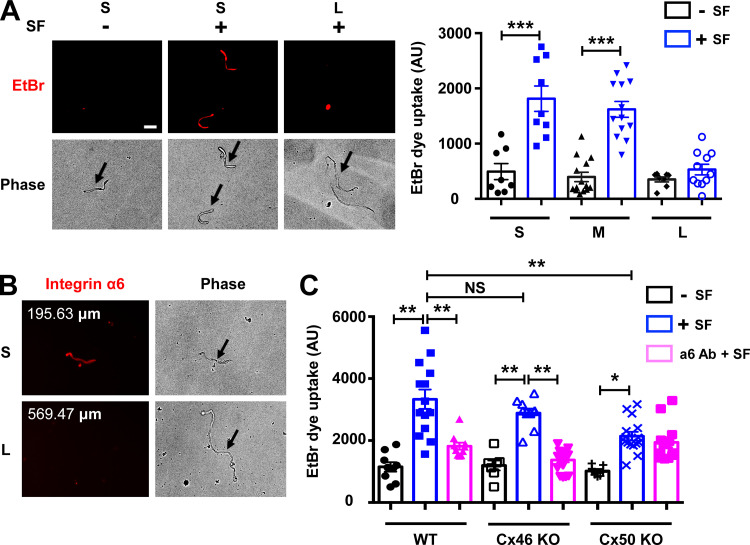
Figure 8.
Integrin α6 is required for Cx50 hemichannel opening. (A) Different lengths of single lens fiber cells were isolated from WT mice and mechanically loaded by spinning force (SF; 1,000 rpm for 5 min). An EtBr dye uptake assay was performed. Scale bar, 50 µm (left). The morphology of unhealthy fiber cells resembles resealed globules or has a rounded appearance (Bhatnagar et al., 1995). Unhealthy or dead fibers are readily distinguished by their morphology without using FITC dextran. Isolated single fibers were grouped by their lengths; short fibers (S) were <200 µm long; medium fibers (M) were 200 to 400 µm; and long fibers (L) were >400 µm. EtBr dye uptake intensity was quantified by ImageJ (right). (B) Isolated single lens fibers were obtained from WT mouse lenses and immunostained with integrin α6 antibody. (C) Single lens fibers were isolated from WT, Cx46 KO, and Cx50 KO mouse lenses. The fibers were pretreated with or without anti-integrin α6 antibody (α6 Ab; 10 µg/ml for 20 min), and EtBr dye uptake assay was performed during mechanical stimulation with SF (1,000 rpm for 5 min). EtBr dye uptake intensity in short fibers was quantified. Each data point in the graph represents an individual single fiber in one of three independent experiments. All data are presented as mean ± SEM. NS, not significant; *, P < 0.05; **, P < 0.01; ***, P < 0.001.

