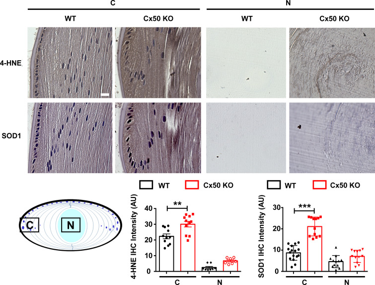
Figure S5.
Increased oxidative stress in the Cx50-deficient mouse lens. Immunohistochemical staining with anti-4-HNE and anti-SOD1 antibodies was adopted to assess the oxidative stress level in WT and Cx50 KO mice lens. Sagittal paraffin sections of 2-mo-old mouse lenses from WT and Cx50 KO mice were prepared. The ABC (avidin–biotin–peroxidase complex) Immunostaining Assay Kit (PK-6101; Vector Laboratories) was used. Briefly, lens tissue sections were antigen unmasked using sodium citrate buffer (pH 6.0) at 65°C for 2 h for 4-HNE and SOD1 staining. Lens tissue sections were then treated with rabbit normal serum for 20 min at room temperature to block nonspecific staining. Tissue sections were stained with anti-4-HNE monoclonal antibody (1:150 dilution; ab46545; Abcam) or anti-SOD1 antibody (1:200 dilution; SC11407; Santa Cruz) for 30 min at room temperature. The sections were then incubated for 30 min with biotin-labeled secondary antibody and VECTASTAIN ABC Reagent for 30 min. Samples were washed in PBS and developed in DAB (SK4100) chromogen solution (Vector Laboratories). Tissues were then counterstained with Hematoxylin (H-3401; Vector Laboratories) for 5 min at room temperature and mounted. Sections were photographed using a Keyence microscope (BZ-X710). The expression level of 4-HNE and SOD1 (brown signals, upper panel) in the cortical lens (C) and nuclear lens (N) was quantified using ImageJ, and reciprocal intensity (Nguyen et al., 2013) is displayed (lower panel). Each data point in the graph represents an individual quantified region in one of three independent experiments. Scale bar, 20 µm. The data are presented as mean ± SEM. **, P < 0.01; ***, P < 0.001.

