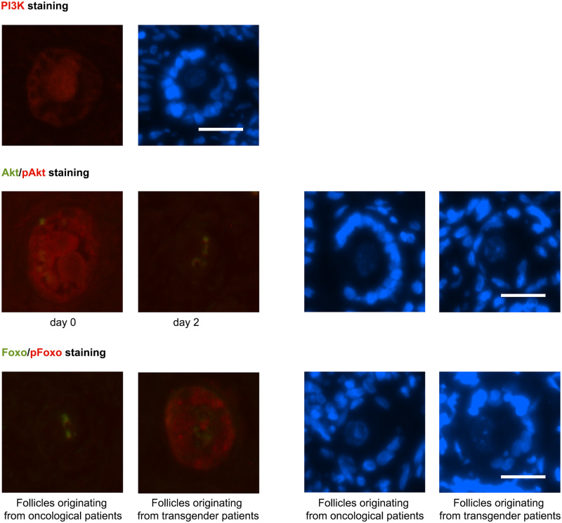
Figure 7.
IHC PI3K Akt pathway. Upper panel: PI3K was seen in the cytoplasm of primordial, intermediate and primary oocytes and in all granulosa cells (red dye, oncological primary follicle culture Day 4). Middle panel: Akt (green dye) and pAkt (red dye) (oncological intermediate follicles on culture Day 0 and 2). Akt was localised as a hotspot in the oocytes cytoplasm of primordial, intermediate and primary follicles from Day 0 until Day 6. The active counterpart, phosphorylated Akt (pAkt), was diffusely located in the entire oocyte cytoplasm on Day 0 in primordial, intermediate and primary follicles. This diffuse localisation reorganised into a cytoplasmic hotspot on later culture days, independent of the follicle stage. Lower panel: FOXO1 (green dye) and pFOXO1 (red dye) (intermediate follicles culture Day 2 from oncological patients and transgender men). FOXO1 was seen as a hotspot in the oocyte in primordial, intermediate and primary follicles, from Day 0 until Day 4. The inactive phosphorylated 1 (pFOXO1) was observed in both oocyte and granulosa cells. In granulosa cells, pFOXO1 could not be found in from oncological patients derived primordial granulosa cells on every culture day and in intermediate granulosa cells on Day 0 and 2 compared to pFOXO presence in all other conditions. The right panel of the picture pairs represents the same follicles stained with DAPI as a reference. Scale bar = 25 µM.

