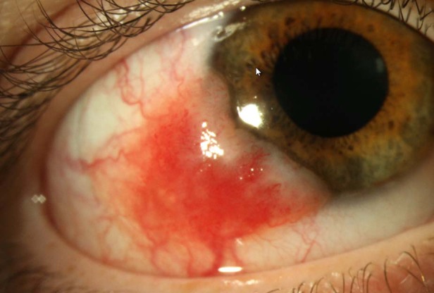
Fig. 2. Photograph of the naevus that presented with features suspicous of malignancy.

These features were: largest basal diameter 9mm, corneal involvement, feeder vessels and recurrence at the site of a previously excised atypical naevus.

These features were: largest basal diameter 9mm, corneal involvement, feeder vessels and recurrence at the site of a previously excised atypical naevus.