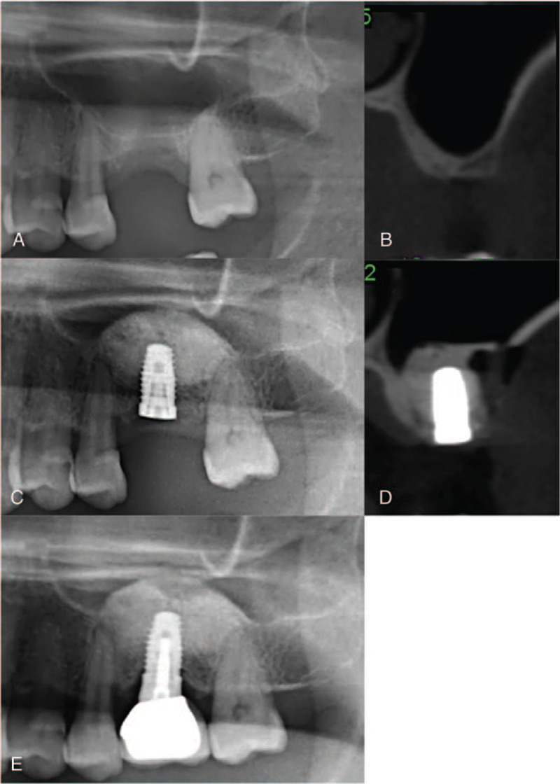
Figure 3.

Representative CBCT radiographs demonstrate procedural steps for the 1-stage lateral window sinus lift (L-1 group). (A, B) Pre-operative radiographs show extremely atrophic residual bone height (RBH) at upper left first molar region (RBH: 2 mm). (C, D) Radiographs obtained immediately after lateral window sinus lift with simultaneous implant placement. (E) Radiograph obtained 18 months after prosthesis delivery.
