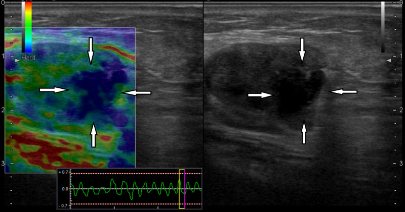
Figure 1. A 77-year-old male with a diagnosis of malignant melanoma of the left ankle.
In the left inguinal lymph node longitudinal sonogram, the lymph node had the heterogeneous round hypoechoic appearance and lobulated contour with irregularity in the B-mode examination (white arrows on right). In the elastography examination (left), there were metastatic hard areas with extension beyond the contour (white arrows on left). Histopathologically, the lymph node was diagnosed as metastatic with pericapsular spread

