Figure 3.

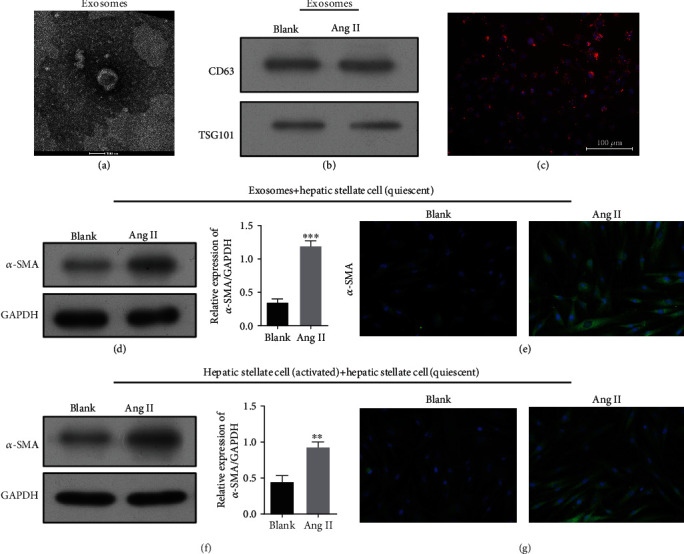
ERS caused exosome release from activated HSC cells. Exosomes released by LX-2 cells in the Ang II and blank groups were detected by electron microscopy (a). (b) Western blot analysis of α-SMA expression in exosomes released by LX-2 cells in the Ang II and blank groups. PKH-26-labeled exosomes (red) were taken up by LX-2 cells (c). Scale bar, 100 nm. Western blotting (d) and immunofluorescence staining (e) of α-SMA in quiescent HSC cells after incubated with exosomes released from activated HSC cells. Western blotting (f) and immunofluoresce nce staining (g) of α-SMA in quiescent HSC cells cocultured with activated HSCs. ∗∗p < 0.01 and ∗∗∗p < 0.001 vs. blank.
