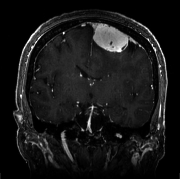
FIGURE 2.

Brain magnetic resonance imaging.Brain magnetic resonance imaging revealed a dural‐based mass of 3 cm in diameter in the left parietal lobe. This mass was removed by surgery and a diagnosis of meningioma was made on the basis of pathological examination findings
