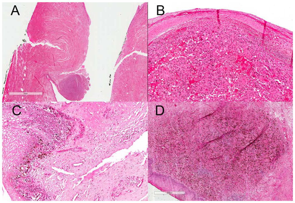
Figure 1:
A : Scanning magnification view (3X view) of VMM showing atypical epitheliod cells (melanocytes) in epidermis on H&E slide; Higher magnification (300X view, B & C and 600X view, D) of VMM showing atypical epitheliod cells (melanocytes) in epidermis and dermis with scattered melanin pigments in dermis on H&E slide
Note: The glass slide is almost 34 years old, and there has been some fading over time.

