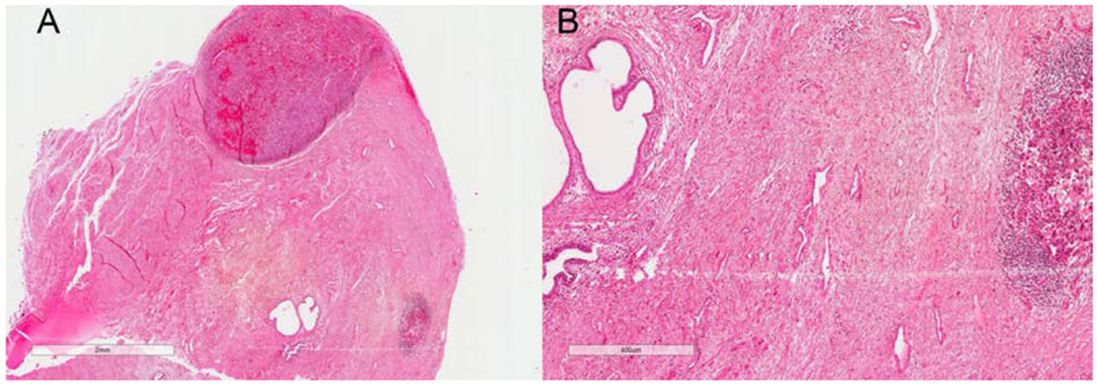
Figure 2:
A: Scanning magnification view (2X view) of VMM showing periurethral involvement by VMM and dermal boundaries in relation to Clark level of invasion and tumor thickness on H&E slide; B: Higher magnification view (400X view) of periurethral involvement by VMM and atypical epitheliod cells (melanocytes) in dermis on H&E slide
Note: The glass slide is almost 34 years old, and there has been some fading over time.

