Figure 3.

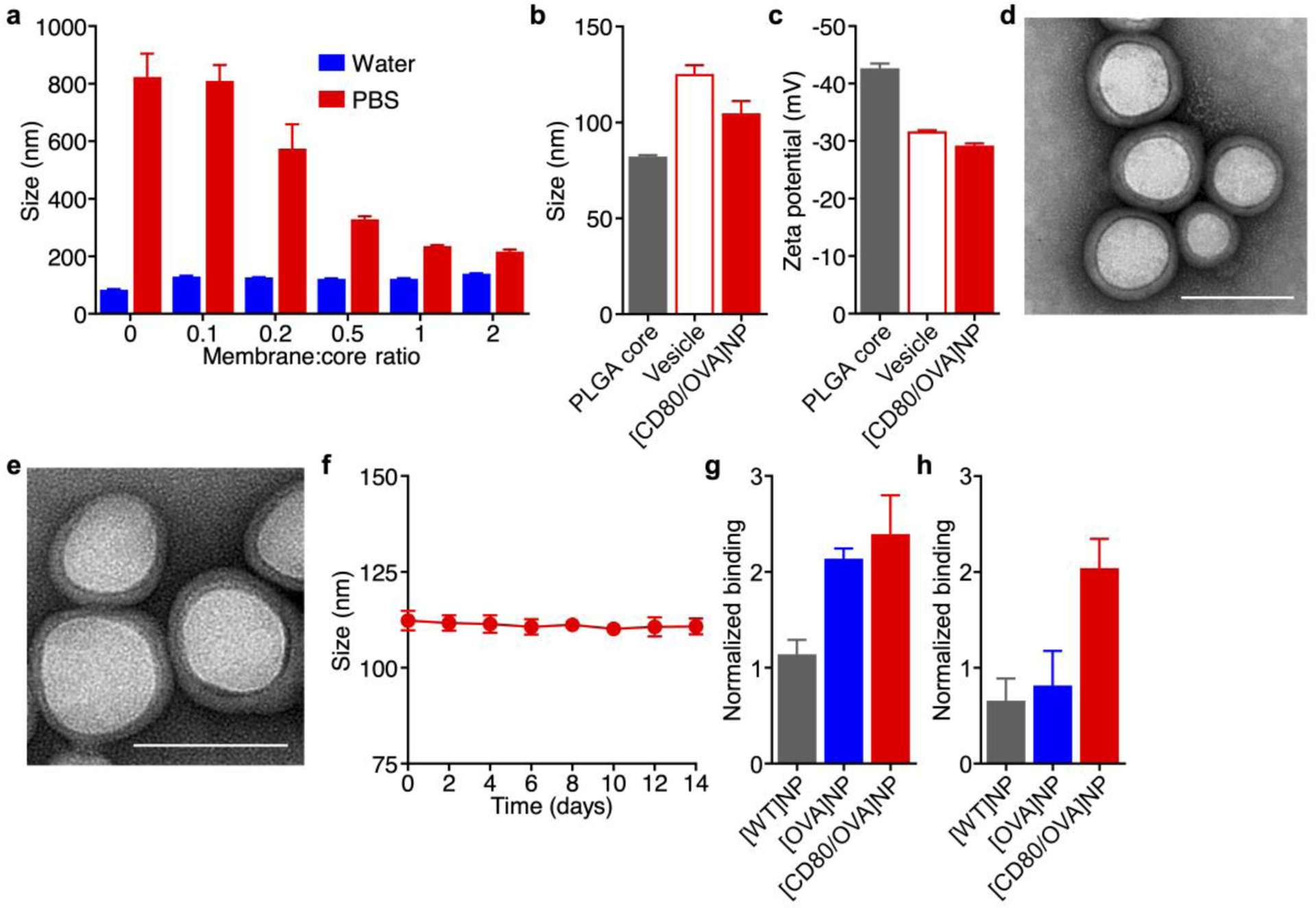
Fabrication and characterization of engineered antigen-presenting nanoparticles. a) Size of [CD80/OVA]NPs at different membrane to core weight ratios when suspended in water or PBS (n = 3; mean + SD). b,c) Hydrodynamic diameter (b) and surface zeta potential (c) of bare PLGA cores, B16-CD80/OVA membrane vesicles, and [CD80/OVA]NPs (n = 3; mean + SD). d,e) Transmission electron microscope images of [CD80/OVA]NPs immediately after synthesis (d) and after 1 week of storage (e). Scale bars = 100 nm. f) Size of [CD80/OVA]NPs over 2 weeks (n = 3; mean ± SD). g,h) Relative binding of antibodies against Kb-SIINFEKL (g) and CD80 (h) to [WT]NPs, [OVA]NPs, and [CD80/OVA]NPs (n = 3; mean + SD).
