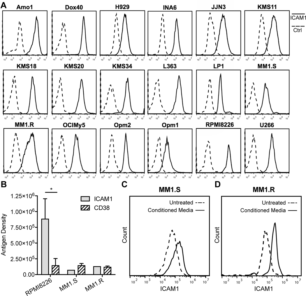
Figure 1. Assessing ICAM1 expression in myeloma cell lines by flow cytometry.
(A) Histograms showing the anti-ICAM1 IgG1 M10A12 binding (solid lines) on the cell surface of 18 myeloma cell lines, compared to a nonbinding IgG1 (Ctrl, dashed lines). (B) Cell surface antigen density of ICAM1 compared to CD38 for representative myeloma cell lines RPMI8226, MM1.S and MM1.R. Student t-test, unpaired, two tailed. *, P < 0.05. (C & D) ICAM1 cell surface expression is increased in myeloma cell lines MM1.S (C) and MM1.R (D) when incubated with HS5 conditioned media (CM) for 3 days compared to equivalent serum conditions without conditioned media (untreated).

