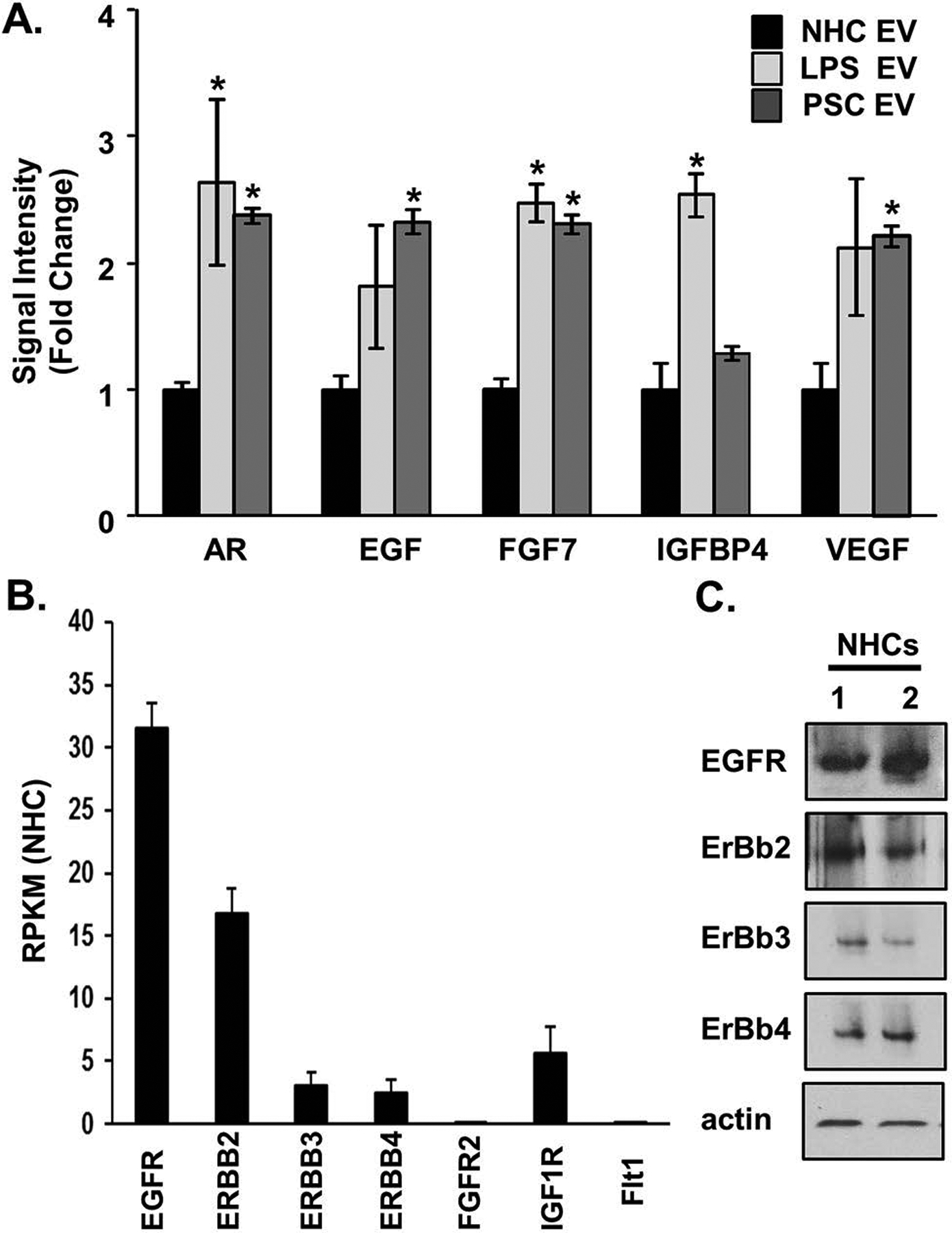
Figure 3. Senescent cholangiocyte-derived extracellular vesicles (EVs) are enriched in growth factors.

A, Equal numbers of normal and senescent cholangiocyte-derived EVs were harvested for protein analysis using a growth factor array (Abcam). Bars represent mean (±) standard error of the mean (SEM); n=3.The primary sclerosing cholangitis (PSC) values represent the mean of 3 PSC patient samples. Values represent a fold change versus EVs derived from normal human cholangiocytes (NHCs). *P<0.05. B, RNA-sequencing data on NHCs, shown as reads per Kilobase of transcript, per million mapped reads (RPKM) from 3 biological replicates revealed increased expression of the epidermal growth factor receptors (EGFRs), EGFR and Erb-B2 receptor tyrosine kinase 2 (ERBB2), compared to the fibroblast growth factor receptor (FGFR2), the insulin-like growth factor 1 receptor (IGF1R), and FMS-related tyrosine kinase 1 (FLT1, ie, VEGFR-1). C, Immunoblotting analysis on NHCs (2 biological replicates) showed positive protein expression of the EGF receptors.
