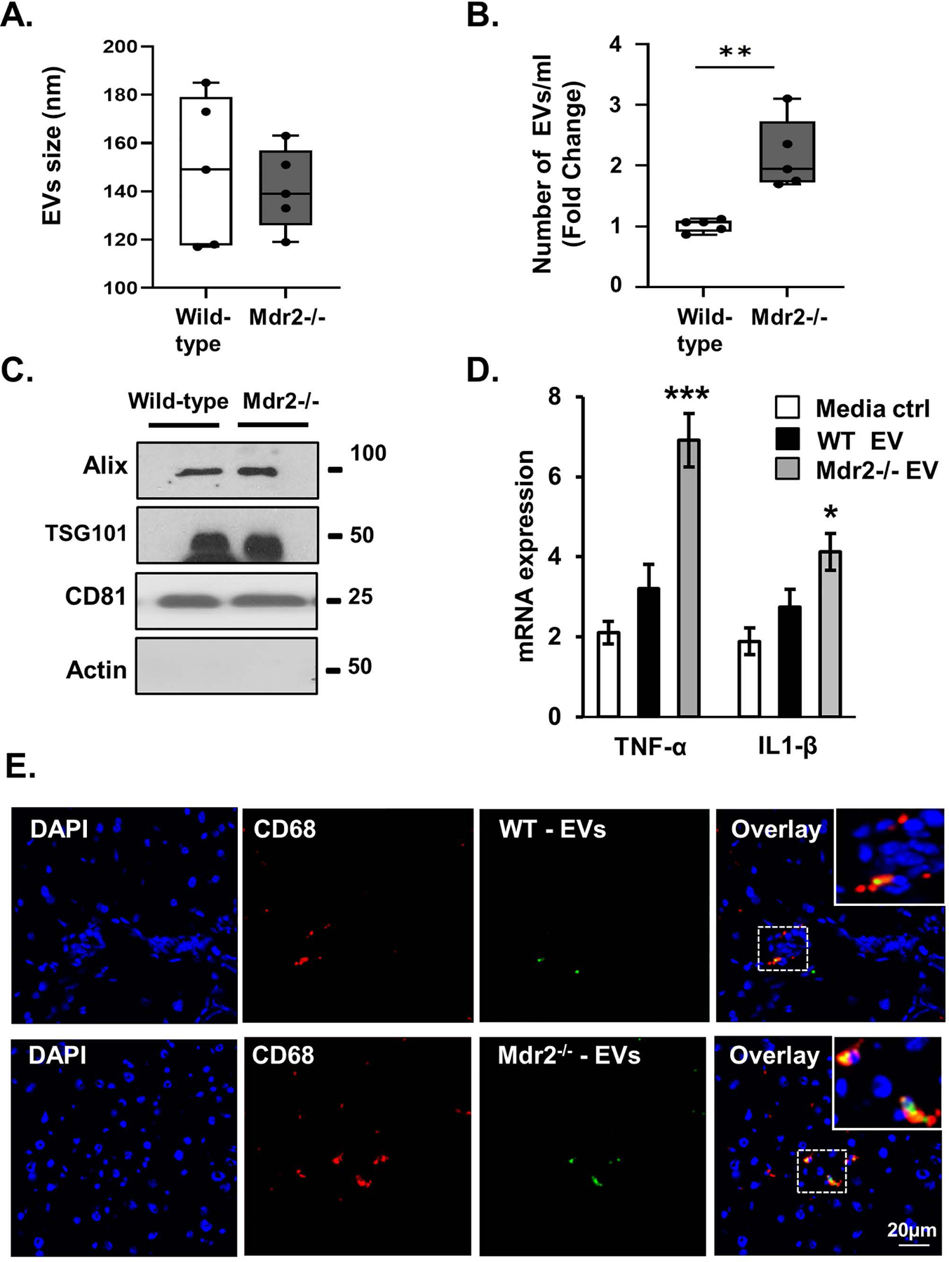
Figure 7. Circulating extracellular vesicles (EVs) are increased in the Mdr2−/− murine model of primary sclerosing cholangitis (PSC).

A and B, Characterization of wild-type C57BL/6J and Mdr2−/− mouse plasma-derived EVs, vesicle size, and number using nanoparticle tracking analysis (NTA) S300. The EVs isolated from wild-type and Mdr2−/− were similar size, yet more EVs were detected in the plasma of Mdr2−/− mice compared to wild-type mice. Data represent size and number from 5 mice per group. C, Western blotting analysis indicated that wild-type and Mdr2−/− mouse-derived EVs were positive for the exosome marker Alix, TSG101, and CD81. D, Equal numbers of wild-type and Mdr2−/− mouse-derived EVs (1e8/ml) were applied to a mouse monocyte cell line (RAW). Activation after 24-hours incubation was assessed by measuring monocyte messenger RNA (mRNA) expression levels (RT-qPCR) of tumor necrosis factor (Tnf) and interleukin 1 beta (Il1b). E, In vivo EV uptake assay reveals that Dio dye-labeled plasma-derived EVs colocalize with hepatic monocytes. EVs were collected from wild-type (WT) or Mdr2−/− mouse plasma and labeled with DiO dye. Immunofluorescence reveals colocalization of CD68 (red, monocytes marker) and plasma EVs (green, labeled with DiO dye) in liver tissue of WT (upper panel) and Mdr2−/− (lower panel) mice. Bars represent mean (±) standard error of the mean (SEM); n=3. *P<0.05, **P<0.01.
