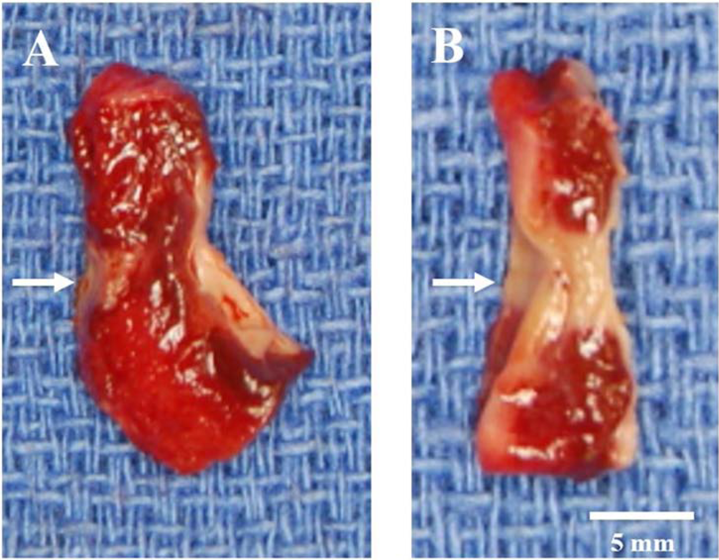
Figure 3.

Cross-section samples of non-transmural (A) and transmural (B) ablations after TTC staining. The ablation line is indicated by arrows. Devitalized tissue does not take up TTC, thus the ablations appear white or pale. The non-transmural ablated tissue is indicated by a red color. The endocardial surface is on the right side of the tissue samples.
